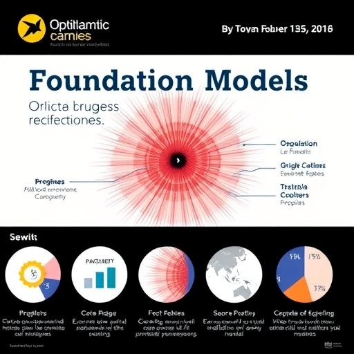
Artificial intelligence is moving ophthalmology toward a new era in which algorithms may analyze not only retinal photographs, but also optical coherence tomography, scanning laser ophthalmoscopy, clinical records, and other forms of patient data in a single diagnostic framework. A review published in Eye & ENT Research describes how ophthalmic foundation models are being developed to perform this kind of multimodal reasoning, while warning that impressive laboratory results have not yet translated into sufficient real-world evidence.
Traditional convolutional neural networks, or CNNs, have already achieved strong performance in narrowly defined ophthalmic tasks. These systems learn to identify patterns associated with diseases such as diabetic retinopathy, glaucoma, and age-related macular degeneration. However, conventional CNNs generally require large collections of images labeled by expert clinicians, and they are usually designed for one specific task or imaging modality. That limits their ability to generalize to new hospitals, populations, diseases, or combinations of clinical evidence.
Foundation models attempt to overcome these constraints through self-supervised learning. Instead of relying exclusively on manually labeled images, they are trained on very large datasets in which the model learns the underlying structure of the data by solving prediction tasks. In ophthalmology, this can involve reconstructing missing portions of images, matching different views of the same eye, or learning relationships between images and clinical descriptions. The resulting representations can then be adapted to multiple diagnostic tasks with relatively little additional labeled data, a capability known as few-shot learning. In some cases, the models can also attempt zero-shot prediction, applying knowledge to a task for which they received little or no task-specific training.
The review traces a rapid progression of these systems. RETFound demonstrated that self-supervised pretraining could produce useful and generalizable representations for retinal imaging, although it was not designed to fuse multiple data types. VisionFM expanded the concept with a modality-agnostic decoder capable of supporting eight ophthalmic imaging modalities. EyeCLIP brought together 11 imaging modalities and clinical text, allowing visual information to be linked with language-based medical knowledge. EyeFM combined five imaging modalities with a large language model, while MIRAGE concentrated on paired optical coherence tomography and scanning laser ophthalmoscopy data. Across reported evaluations, multimodal systems generally outperformed comparable single-modality models, particularly when only a small number of labeled examples were available.
The technical advantage of multimodal fusion is that different forms of evidence can capture different aspects of disease. A retinal photograph may reveal vascular abnormalities, an OCT scan can show microscopic changes in retinal layers, and clinical text may provide information about symptoms, medical history, medications, or systemic risk factors. A model that processes these inputs jointly can potentially learn relationships that are invisible when each source is analyzed separately. Yet the review emphasizes that combining modalities is not equivalent to reproducing clinical reasoning. The model may identify statistical patterns across data sources without understanding disease mechanisms or establishing why a particular finding matters.
The most substantial clinical evidence currently comes from EyeFM. In a double-blind randomized controlled trial, physicians supported by the AI system achieved a diagnostic accuracy of 92.2 percent, compared with 75.4 percent among physicians working independently. The AI-assisted group also reduced report-writing time by an average of 63.3 seconds. The result is notable because it evaluates the system in a clinical workflow rather than only on a retrospective image archive. It is also, according to the review, the only randomized controlled trial-based validation reported among the five foundation models discussed.
The contrast with the rest of the field is considerable. RETFound, VisionFM, EyeCLIP, and MIRAGE have been evaluated exclusively using retrospective datasets, and the review states that these models lack prospective, multicenter validation. Retrospective studies can reveal whether an algorithm recognizes patterns in previously collected data, but they may overestimate performance because of hidden biases, differences in image quality, or overlap between training and evaluation populations. Prospective studies conducted across hospitals are needed to determine whether these systems remain reliable when confronted with new equipment, diverse patients, incomplete records, and the time pressures of routine care.
Some of the models have also been tested for clues to systemic disease hidden in ophthalmic images. RETFound produced area-under-the-receiver-operating-characteristic values of 0.737 for myocardial infarction, 0.794 for heart failure, and 0.754 for ischemic stroke prediction. The AUROC is a statistical measure of how well a model separates people with and without a condition; a value of 0.5 corresponds to chance performance, while 1.0 represents perfect discrimination. VisionFM, meanwhile, estimated 38 systemic biomarkers from ophthalmic images with a mean accuracy of 78.6 percent. The review presents these findings as dataset-specific associations, not evidence that eye images causally determine cardiovascular or other systemic outcomes.
Important weaknesses remain beneath the headline numbers. Training datasets often lack adequate demographic diversity, raising concerns that performance may vary across ethnicities, age groups, socioeconomic backgrounds, and healthcare settings. Many systems rely on two-dimensional OCT slices rather than complete three-dimensional volumetric scans, potentially discarding clinically relevant structural information. Interpretability methods, including heat maps and other post-hoc explanations, can indicate which image regions influenced a prediction, but they do not establish a causal mechanism or prove that the model used medically meaningful features. The review therefore argues that the field’s central challenge is no longer simply improving benchmark scores. The decisive test will be whether ophthalmic foundation models can be integrated safely into clinical practice through transparent, prospective, and multicenter evaluation.
The work, titled “Foundation Models in Diagnosis of Ophthalmic Diseases: Construction and Application,” was published in Eye & ENT Research on June 4, 2026. Its authors frame foundation models as a potential bridge between image analysis and broader medical decision-making, but not as replacements for clinicians. Their long-term value will depend on whether they can support physicians consistently, explain their limitations, protect patient data, and perform equitably across real-world populations. For now, the technology’s viral appeal is racing ahead of its clinical evidence, making rigorous validation the essential next step.
Subject of Research: Ophthalmic foundation models and their applications in the diagnosis of eye diseases and prediction of systemic health conditions.
Article Title: “Foundation Models in Diagnosis of Ophthalmic Diseases: Construction and Application”
News Publication Date: Not provided.
Web References: https://doi.org/10.1002/eer3.70041
References: “Foundation Models in Diagnosis of Ophthalmic Diseases: Construction and Application,” Eye & ENT Research, published June 4, 2026. DOI: 10.1002/eer3.70041.
Image Credits: Higher Education Press
Keywords: ophthalmic foundation models, artificial intelligence, ophthalmology, retinal imaging, OCT, multimodal learning, EyeFM, RETFound, VisionFM, EyeCLIP, MIRAGE, clinical validation, medical AI, systemic disease prediction
Tags: artificial intelligence in retinal disease diagnosischallenges in real-world implementation of ophthalmic AIclinical evidence for AI in ophthalmologyconvolutional neural networks for eye disease detectiondeep learning for ophthalmic imagingdiagnostic frameworks combining OCT and fundus imagingfuture directions in ophthalmic AI developmentlaboratory versus clinical performance of ophthalmic modelsmachine learning for glaucoma and macular degenerationmultimodal medical data analysis in ophthalmologyOphthalmic foundation modelsself-supervised learning in ophthalmology