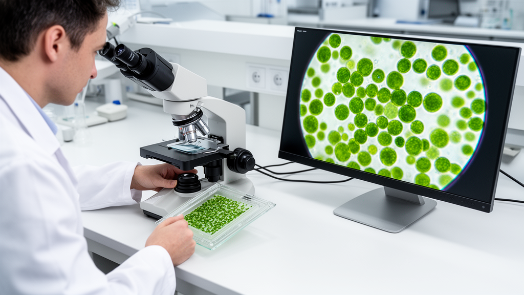
Accurate quantification of microalgal cell density is essential for both basic biological research and commercial applications because cell counts directly indicate culture growth stages, overall productivity, and physiological status. Although density measurements underpin operations in sectors such as biofuels, cosmetics, nutraceuticals, and pharmaceuticals, conventional cell counting techniques often require researchers to navigate steep trade-offs between procedural expense, workflow duration, and human error. To resolve these operational bottlenecks, researchers developed CellScore, an open-source, automated image-processing pipeline implemented within the widely used biological image analysis platform ImageJ. The pipeline pairs standard hemocytometer preparation with automated digital analysis, allowing laboratories to achieve rapid cell counts from routine microscopy without relying on specialized commercial devices or high-cost instruments.
Traditional hemocytometry remains widely accessible across scientific institutions because it demands only a light microscope, a hemocytometer slide, and basic accessories, delivering high accuracy when performed carefully. However, manual counting requires operators to track and tally each individual cell, consuming significant time and introducing human error that fluctuates based on user expertise and fatigue. While spectrophotometric optical density measurements provide a rapid alternative by estimating cell numbers via absorbance calibrations, optical density readings at standard wavelengths such as 680 nanometers or 750 nanometers introduce distinct analytical challenges. Absorbance signals vary according to chlorophyll content, environmental stress, or non-algal particulate interference, necessitating repeated empirical verification. Similarly, automated flow cytometry and dedicated benchtop cell counters offer high throughput but carry steep capital costs, require specialized consumable slides, demand extensive protocol optimization for varying cell sizes, and have historically been optimized for larger mammalian cells rather than small microalgal species.
To evaluate CellScore against standard laboratory workflows, the research team examined three microalgae species encompassing distinct genetic traits: six strains of Chlamydomonas reinhardtii, comprising wild-type 21 gr, wall-deficient cw15, wall-deficient starchless mutants sta6 and sta7, and flagellar mutants pf18- and fla3-1; one strain of Chlorella sorokiniana KAS908; and one culture of Micractinium reisseri PRAS. Cultures were grown in liquid tris-acetate phosphate medium under continuous agitation at 120 revolutions per minute with a light intensity of 80 micromoles per square meter per second for two days, receiving fresh medium every two days for one week. Culture samples were harvested after 48 to 72 hours of growth once spectrophotometric absorbance measurements plateaued at stationary phase. Unstandardized brightfield images of Neubauer hemocytometer grids loaded with 10 microliters of sample at specified dilutions were subsequently captured at 10-times magnification using a Nikon ECLIPSE Si trinocular microscope paired with a RisingCam digital camera.
The CellScore workflow operates by leveraging ImageJ’s native Analyze Particles tool, preceded by an automated sequence of digital pre-processing routines. The software first converts imported color images into greyscale format and executes binarization using a manually selected threshold range to separate cells from the background field. To counteract artifacts created during aggressive thresholding, the macro automatically fills holes within identified cellular structures and executes binary watershed operations to segregate contiguous or overlapping cells. Operators calibrate the macro by defining three primary user parameters: the intensity threshold range, cell size boundaries, and circularity values. Because circularity is mathematically defined as four times pi multiplied by area divided by perimeter squared, users outline representative cells using ImageJ’s freehand tool to obtain preliminary shape descriptors and area measurements, adjusting baseline boundaries slightly to encompass natural biological variation within the culture.
The investigative team compared CellScore’s performance directly against three alternate counting methodologies across an identical evaluation set of 36 hemocytometer quadrant images: manual counting using a physical clicker, automated counting utilizing a dual-software pipeline of Ilastik segmentation paired with CellProfiler quantification, and automated counting via ImageJ’s unassisted Analyze Particles function. Ground-truth control values were established by manually tallying each individual cell using the ImageJ Cell Counter plugin. For C. reinhardtii 21 gr, manual ImageJ counting yielded an average of 276 cells per image over 21 minutes, while the manual clicker recorded an average of 295 cells in 9 minutes with a 7.0 percent error rate. In contrast, CellScore processed the 21 gr images in approximately 2.2 minutes, recording an average of 280 cells with an error rate of only 1.3 percent relative to the manual control.
Across the diverse microalgal strains and mutants evaluated, CellScore maintained consistent counting accuracy while substantially reducing processing durations compared to manual workflows. In evaluations of the wall-deficient mutant C. reinhardtii cw15, CellScore required 1.7 minutes to identify an average of 123 cells with 2.9 percent error, whereas the manual clicker required 4 minutes with 3.1 percent error. High-density cultures demonstrated even greater absolute time savings. For C. sorokiniana KAS908, where manual ImageJ counting required 186 minutes to register an average of 3,258 cells and manual clicker counting took 105 minutes with 9.0 percent error, CellScore tallied an average of 3,194 cells in just 1.6 minutes with a 2.0 percent error. In dense cultures of M. reisseri PRAS averaging 1,934 cells by manual ImageJ counting over 80 minutes, CellScore recorded an average of 1,965 cells in approximately 5 minutes with a 1.6 percent error rate, outperforming clicker counts that showed an 8.1 percent error over an hour of manual effort.
Statistical evaluation confirmed that CellScore delivers reliable counts without introducing systematic bias. A two-tailed Wilcoxon signed-rank test conducted at an alpha threshold of 0.01 evaluated the differences between the manual ImageJ control and each experimental counting technique across the 36 test images. The analysis revealed that the manual clicker, the Ilastik-CellProfiler pipeline, and the automated CellScore method each yielded test statistics exceeding the critical threshold, failing to reject the null hypothesis and demonstrating no statistically significant difference from the manual control. In contrast, the unassisted ImageJ Analyze Particles method produced a test statistic smaller than the critical value, rejecting the null hypothesis and confirming statistically significant deviation from ground-truth counts. The poor performance of unassisted ImageJ counting, which exhibited error rates as high as 29.7 percent on wild-type C. reinhardtii, was attributed to the absence of crucial pre-processing steps like hole filling and watershed segmentation, leaving the native algorithm vulnerable to background noise and cell clumping.
To assess software accessibility and usability for untrained personnel, the authors tested CellScore with two novice users who possessed no prior experience operating the pipeline. Across identical image sets, both novice operators achieved cell counts with error rates remaining below 15 percent, requiring less than an average of eight minutes of initial setup time per image and under one minute of active execution time. Furthermore, independent verification using automated overlay scripts developed in MATLAB and Python demonstrated that CellScore produced an average of six false-positive detections and five false-negative classifications per 100 counted cells. The authors observed that the majority of false-positive detections arose from thresholding artifacts that mistakenly marked background hemocytometer grid lines as cellular bodies, an issue that can be minimized through precise threshold calibration or by analyzing cell suspensions on plain, non-gridded glass slides when absolute volumetric concentrations are not strictly required.
The researchers noted specific technical limitations in the present pipeline that establish clear boundaries for its current application. Although tested across multiple strains and cell-wall variants, all three examined species—Chlamydomonas reinhardtii, Chlorella sorokiniana, and Micractinium reisseri—share relatively similar spherical or ellipsoidal morphologies, meaning that broader calibration and validation remain necessary before deploying CellScore on organisms with irregular shapes, elongated filaments, or complex colonial structures. Looking forward, the authors highlighted opportunities to expand the open-source pipeline by integrating automated viability sorting to distinguish live from trypan blue-stained dead cells, adapting detection parameters for confocal fluorescence and chlorophyll autofluorescence microscopy, and testing performance on other industrially relevant microorganisms such as bacteria and yeasts.
Subject of Research: Automated image-based microalgal cell quantification using open-source ImageJ software
Article Title: CellScore: an open-source image-based pipeline for automated cell quantification in microalgae
Article References: Do, S., Huang, S., Plan, A., Birn, N., Walker, A., & Du, Z.-Y. (2026). CellScore: an open-source image-based pipeline for automated cell quantification in microalgae. Advanced Biotechnology, 4(4), 46. https://doi.org/10.1007/s44307-026-00131-w
Image Credits: AI Generated
DOI: 10.1007/s44307-026-00131-w
Keywords: microalgae, CellScore, ImageJ, cell counting, hemocytometer, bioimaging, biotechnology, Chlamydomonas reinhardtii, open-source, image-based, pipeline, automated
News Source: Gregory Coleman. (November 9, 2026). Open-Source CellScore Pipeline Automates Microalgal Cell Counting Using Standard Microscopy. Scienmag.