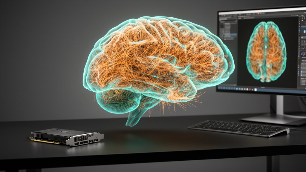
A single mouse brain imaged with modern light-sheet microscopy or serial two-photon tomography produces a torrent of numbers that most laboratories cannot even open, let alone interpret. Volumetric datasets routinely reach from several terabytes to more than ten terabytes, and while imaging throughput has accelerated dramatically in recent years, converting those raw voxels into biological meaning—cell counts, axonal lengths, vascular branch densities—has remained the true bottleneck of whole-brain neuroscience. A team of researchers in China now reports a software package designed to collapse that entire bottleneck into one window on an ordinary desktop computer.
The tool, called BVLab-Analyzer, is described in a study published in Frontiers in Neuroinformatics by Ying Hu of Hubei University of Education, Xiaojun Wang of Hainan University, and colleagues at the Britton Chance Center for Biomedical Photonics at Huazhong University of Science and Technology. It is a standalone, zero-code graphical application for Windows that carries a terabyte-scale brain dataset through every stage of analysis: importing data, annotating training examples, training a deep neural network, running whole-brain inference, stitching results together, and exporting standardized morphological reconstructions and statistics. Crucially, the entire workflow runs on a single consumer-grade graphics card.
That last point is more radical than it may sound. Most existing deep-learning frameworks for whole-brain analysis—Cellfinder, nnU-Net-based pipelines, and others—assume access to multi-GPU clusters or large-memory enterprise servers. For a typical biology laboratory equipped with one workstation and a gaming-class GPU, whole-brain quantification has effectively been out of reach. BVLab-Analyzer tackles the hardware problem with an adaptive 3D block-wise tiling scheme: the software automatically slices a complete brain volume, which can span three to thirteen terabytes, into tens of thousands of independent sub-volume blocks sized to fit within 8 to 14 gigabytes of GPU memory. Cell detection uses anisotropic blocks of 272 by 272 by 144 voxels to capture the wide footprint of neuronal cell bodies, while neuron and vessel tracing use isotropic 192-cubed blocks to preserve continuity along fibers and vessels in depth.
The architecture itself is a hybrid of C++ for compute-intensive operations and Python for the deep-learning logic, wrapped in a Qt graphical interface and compiled into a pre-bundled executable that eliminates the notorious pain of configuring Python environments and CUDA versions. Data flows through six tabbed modules—Data I/O, Annotation, Training, Inference, Post-processing, and Statistics—all coordinated by a central project file that preserves parameters and fault-tolerant state. The software reads multi-page TIFF stacks, the BioimageVision format, and the cloud-optimized OME-Zarr format, automatically parsing multi-resolution pyramids so users can move between overview and detail levels during visualization, which is rendered in real time through the VTK library.
Training a deep network normally demands enormous annotated datasets, and here the researchers introduced what may be the software’s most elegant trick. A built-in K-Means clustering algorithm extracts grayscale histogram and maximum-intensity-projection features from every sub-block in a whole brain and groups them into one to two thousand clusters. From each cluster, the block nearest the center is offered as a candidate, and the researcher hand-picks only about one to two hundred representative blocks for sparse annotation. This data-driven sampling reduced the required annotation volume by roughly an order of magnitude compared with experience-based random selection. In the reported experiments, training sets of just 166 blocks for cells, 86 for neurons, and 120 for vessels were enough.
Annotation itself is accelerated by what the authors call Smart Annotation tools. For point-like cell bodies, a single click at the soma center registers a 3D coordinate in under a second and stores it as a root node in the standard SWC topology format. For linear structures such as axons, dendrites, and blood vessels, users place sparse anchor points along the fiber, and the software auto-interpolates the intermediate skeleton path with parent-child links compliant with the SWC specification. Large vessels that appear hollow or faint after tissue clearing get a dedicated ray-casting mode that projects orthogonal rays from reference points, detects the vessel wall boundary by local intensity drops, and computes the true cross-sectional midpoint and radius. Sparse cell points are then converted to Gaussian blobs and skeleton trajectories into tubular Gaussian volumes, producing dense training masks without ever requiring pixel-level contour drawing.
For inference, the trained 3D U-Net sweeps the whole brain with overlapping sliding windows whose overlaps are tuned to biology: 32 by 32 by 20 voxels for cells, roughly one and a half to two soma diameters, and 4 by 4 by 2 for the thin linear structures. After prediction, the topologies of all blocks are merged back into whole-brain coordinate space. Perhaps the most practically beloved feature will be the interrupt-and-resume engine. Whole-brain prediction can take up to 129 hours, and on shared lab workstations interruptions are inevitable. The software logs completed sub-volumes in real time, so after a power outage or a colleague borrowing the machine for the day, it simply skips the finished blocks and continues—validated in the study by pausing a neuron reconstruction after 48 hours, using the workstation for office work, and resuming without any loss of progress.
The performance numbers are competitive with the specialized state of the art. On a 3.6-terabyte, 16-bit c-Fos cell-detection volume, the pipeline achieved a median precision of 0.863 and an F1-score of 0.868, despite grayscale means that stepped from several hundred to nearly 3,000 across the brain. On a 12.36-terabyte GFP-labeled neuron dataset—the largest tested—the median recall reached 0.893 with an F1 of 0.884, maintaining continuous tracing even under ultra-low-contrast fiber signals. On a 0.47-terabyte vascular volume, the median recall hit 0.944 with an F1 of 0.899, though large-vessel luminal cavities remain a known weak spot. Total training time per task was kept between 1.9 and 2.8 hours on a single RTX 4090, with GPU memory stable at 8 to 14 gigabytes throughout.
What truly distinguishes BVLab-Analyzer, the authors argue, is scope. Cell somata, neuronal processes, and the microvasculature are not isolated objects but together form the neurovascular unit, the functional target of studies of neurovascular coupling. Until now, a laboratory wanting to correlate all three had to juggle separate specialized tools—ClearMap, Cellfinder, TrailMap, Vaa3D—with incompatible output formats and manual bridging between pipelines. BVLab-Analyzer unifies all three target structures in one zero-code environment and exports everything in standard SWC format, readable directly in Vaa3D and neuTube, with statistics such as cell density, total fiber length, branch-point counts, bifurcation angles, and vessel diameter distributions exported as CSV tables for immediate plotting.
The team is candid about limitations. The software processes each fluorescence channel with a dedicated model and cannot separate mixed signals within a single channel; it currently handles only static, ex vivo rodent whole-brain data, runs only on Windows, and cross-region and cross-specimen generalization has not yet been formally evaluated. Difficult regions—dense cell clusters, extremely low signal-to-noise fibers, hollow large vessels—still rely on built-in interactive proofreading tools rather than automatic quality detection, though uncertainty estimation and adaptive post-processing are planned. A Python API and cross-platform support are on the roadmap. Even so, the package marks a genuine democratization of whole-brain analysis: the kind of terabyte-scale quantification that once demanded a computing cluster and a programmer now fits inside a single desktop window, a single GPU, and a workflow that a bench biologist can drive entirely by hand.
Subject of Research: A unified zero-code software toolbox for terabyte-scale whole-brain cell detection, neuron reconstruction, and vascular network extraction
Article Title: BVLab-analyzer: a unified visual toolbox for terabyte-scale whole-brain cell detection, neuron reconstruction, and vascular network extraction
Article References: Hu, Y., Wang, X., Sun, P., Shen, Y., & Quan, T. (2026). BVLab-analyzer: a unified visual toolbox for terabyte-scale whole-brain cell detection, neuron reconstruction, and vascular network extraction. Frontiers in Neuroinformatics, 20, Article 1925112. https://doi.org/10.3389/fninf.2026.1925112
Image Credits: AI Generated
DOI: 10.3389/fninf.2026.1925112
Keywords: whole-brain imaging, deep learning, BVLab-Analyzer, 3D U-Net, neuron reconstruction, vascular segmentation, cell detection, light-sheet microscopy, SWC format, neuroinformatics, GPU computing, zero-code software
News Source: Cassandra Pierce. (October 15, 2026). One GPU, One Toolbox: Software Brings Terabyte-Scale Whole-Brain Analysis to Any Lab. Scienmag.