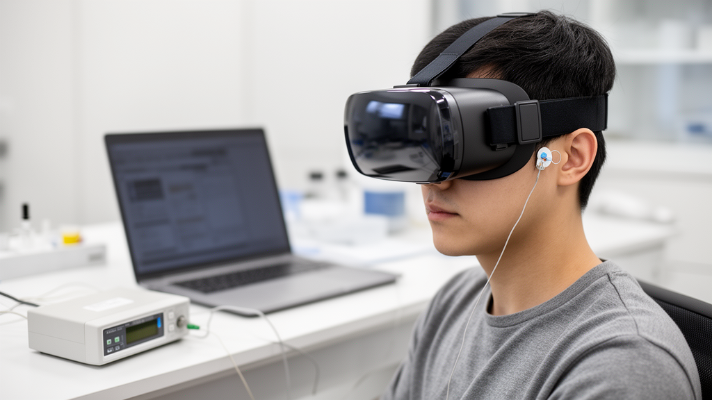
Virtual reality promises immersion, but for many users it delivers something far less pleasant: a creeping wave of nausea, dizziness, and disorientation known as virtual reality motion sickness. The condition, often called cybersickness, arises from a sensory conflict between what the eyes perceive as motion and what the vestibular system in the inner ear reports as stillness. Researchers have long sought objective ways to detect this state before it ruins the experience, and electroencephalography, or EEG, has emerged as one of the most promising tools. Now, a team at Nanjing University of Information Science and Technology has shown that detecting VR motion sickness may not require a skull cap studded with dozens of electrodes. In a study published in Medical & Biological Engineering & Computing, Min Zhu, Chengcheng Hua, and colleagues demonstrate that a single, carefully chosen EEG lead, combined with an interpretable machine learning framework, can classify motion sickness with an accuracy of 87.53 percent.
The motivation behind the work is practical rather than purely scientific. Conventional EEG-based motion sickness detection typically relies on multi-channel recordings and high-dimensional feature sets, which inflate processing complexity, raise device costs, and burden the user with cumbersome hardware. For VR motion sickness detection to ever work in real time inside a consumer headset, the sensing apparatus must be lightweight, comfortable, and computationally cheap. Reducing the number of electrodes from a full cap to a single lead is one of the most direct routes to that goal, but it raises an obvious question: which electrode, of the many possible positions on the scalp, carries the most informative signal about motion sickness? Answering that question blindly, by testing every position with every possible model, would be prohibitively expensive.
The Nanjing team’s solution leans on explainable artificial intelligence. Rather than treating a deep learning model as an inscrutable black box, they used SHAP, or SHapley Additive exPlanations, a technique borrowed from game theory that assigns each input feature a contribution value for the model’s output. SHAP works by treating the model’s prediction as a cooperative game in which each feature is a player, and then calculating how much each player adds to the payout across all possible coalitions. Applied to EEG, SHAP values reveal which electrodes and which signal characteristics the model actually relies on when deciding whether a user is motion sick. This transparency is what allowed the researchers to prune their system down to a single lead without simply guessing.
Specifically, the researchers took six classical deep learning EEG decoding models and used SHAP values to assess the contribution of every recording lead within each model. The candidate electrodes were then ranked by integrating several lines of evidence: the SHAP-based importance scores, the classification performance of each lead when used alone, differences across EEG frequency bands, and the physiological plausibility of the signal source. Out of this analysis, one electrode emerged as the clear frontrunner: FT11, a position over the right temporal region of the scalp. The choice was notable because temporal electrodes sit near muscles involved in facial expression and jaw movement, regions often avoided in EEG research because of contamination concerns, yet FT11 delivered competitive and relatively consistent performance across all six evaluated models.
With the lead selected, the team turned to the feature dimension. Instead of feeding raw waveforms into a heavyweight neural network, they extracted handcrafted features from the single-lead signal, the kind of interpretable quantities that EEG researchers have used for decades: spectral band powers computed via the fast Fourier transform, time-domain statistics such as Hjorth parameters, and ratios between slow and fast oscillations. These features capture well-documented signatures of motion sickness, including shifts in theta and alpha activity that reflect changes in cognitive load and alertness. But even a single lead yields a large menu of candidate features, many of them redundant. To trim the menu, the researchers devised a correlation-SHAP strategy: features that were highly correlated with one another were treated as redundant, and among redundant groups, the feature with the greatest SHAP contribution was retained.
The final classifier was XGBoost, a gradient-boosted decision tree ensemble prized for its efficiency on tabular data. Under a window-level tenfold cross-validation protocol, the compact model achieved an accuracy of 87.53 percent with a standard deviation of 0.73 percent, using just seven selected features. To put that number in context, it was 9.55 percentage points below a model that used all leads and all features, and only 0.69 percentage points below a model that used the single lead with the full feature set. In other words, the feature screening did nearly all of the heavy lifting: once the right electrode was chosen, seven well-chosen features captured almost all of the discriminative information that dozens of features could provide.
The physiological story behind FT11 is intriguing. The temporal region overlies brain areas connected to vestibular processing, and neuroimaging work has implicated the parieto-insular vestibular cortex as a central hub for the sensory conflict that drives motion sickness. Signals recorded near the temporal scalp also reflect activity from brain networks involved in visual-vestibular integration. At the same time, the authors and the broader literature are candid about a caveat: temporal electrodes are prone to contamination from facial muscles such as the temporalis, and electromyographic artifacts can mimic high-frequency EEG activity. The fact that a temporal lead still performed well suggests either that motion sickness information is genuinely strong in that region or that the feature selection process found muscle-related signatures that co-vary with sickness, for example jaw clenching or facial tension that accompany nausea. Disentangling neural from muscular contributions remains an open challenge for the field.
The team was also unusually transparent about the limits of their generalization claims. In an additional post hoc test using leave-one-subject-out validation, in which the model is trained on all participants except one and then tested on the held-out individual, the globally selected configuration showed limited performance on unseen participants. The authors caution that this result should not be read as a fully independent estimate of cross-subject generalization, because the electrode, classifier, and feature set had all been selected using data from the same cohort before the LOSO test was run. This distinction matters enormously for anyone hoping to deploy such a system: a model that excels within a group of users may stumble when faced with a stranger whose EEG patterns differ. Cross-subject robustness remains one of the hardest problems in EEG-based applications, and honest reporting of this gap is a welcome contrast to papers that quietly omit such tests.
Still, the study points toward a future in which VR headsets monitor their users’ brains as unobtrusively as smartwatches monitor heart rates. A single dry electrode embedded in a headset’s padding, paired with a lightweight classifier running on the device’s own processor, could in principle detect the earliest EEG signatures of motion sickness and intervene automatically, perhaps by reducing the field of view, dimming the virtual horizon, or pausing the experience before nausea takes hold. The interpretable framework also offers a reusable recipe for other single-lead EEG applications, a direction already explored in sleep staging, depression screening, and emotion recognition. By showing that explainability tools like SHAP can guide hardware simplification rather than merely decorate a finished model, the Nanjing researchers have made a case that the path to practical brain-aware interfaces runs not through bigger models and denser electrode arrays, but through smarter, more transparent choices about which tiny sliver of neural signal actually matters.
Subject of Research: Single-lead EEG detection of virtual reality motion sickness using interpretable SHAP-based feature selection
Article Title: Single-Lead EEG-based virtual reality motion sickness detection using interpretable feature selection
Article References: Zhu, M., Hua, C., Wang, Q., Fu, J., Xiang, S., Wei, Z., & Liu, J. (2026). Single-Lead EEG-based virtual reality motion sickness detection using interpretable feature selection. Medical & Biological Engineering & Computing. https://doi.org/10.1007/s11517-026-03702-0
Image Credits: AI Generated
DOI: 10.1007/s11517-026-03702-0
Keywords: virtual reality, motion sickness, EEG, SHAP, feature selection, XGBoost, single-lead EEG, cybersickness, machine learning, brain-computer interface, explainable AI, electrode selection
News Source: Cassandra Pierce. (October 19, 2026). One EEG Electrode May Be Enough to Detect VR Motion Sickness. Scienmag.