


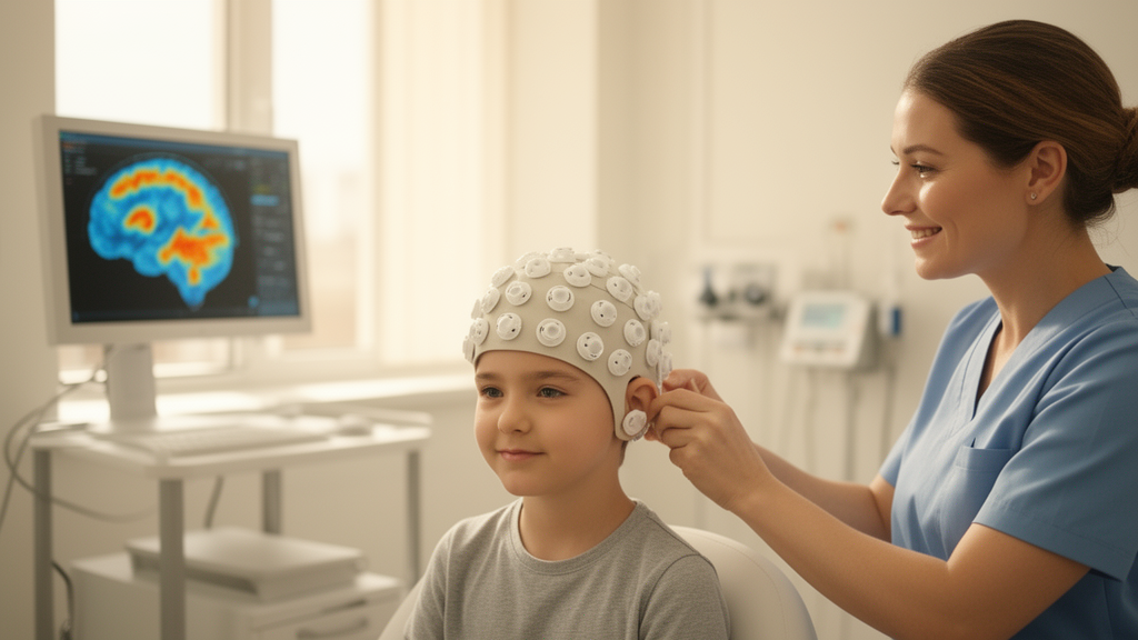
An eight-year-old boy with drug-resistant epilepsy was scheduled for surgery on a lesion sitting squarely in what textbooks call Broca’s area, the classical speech-production region of the left frontal lobe. Cutting there risks taking language with it, and the standard mapping tools were unavailable to him: he could not tolerate the deafening noise of functional MRI and the Wada test was too invasive. Instead, his clinical team turned to a newer technology, magnetoencephalography built from optically pumped magnetometers, worn as a child-sized helmet rather than a rigid cryogenic hood. The measurements reshaped the surgical plan, and the outcome now stands as a detailed single-case demonstration of what on-scalp brain recording can contribute to pediatric epilepsy surgery.
The case, published in Acta Epileptologica, involved a right-handed boy whose seizures began at five years and nine months of age. They started as clapping movements of both hands with repeated blinking, followed by asymmetric stiffening of the upper limbs with deviation to the right and impaired awareness. Before some attacks he felt an aura of numbness in his head and body. The episodes grew relentless, occurring more than ten times a day during both wakefulness and sleep. Oxcarbazepine, levetiracetam, lacosamide, clonazepam and perampanel initially brought them under control, with the longest seizure-free stretch lasting four months, but at six years and five months of age the seizures returned in clusters of one to more than ten per day, this time with awareness preserved, and he was diagnosed with drug-resistant epilepsy.
His presurgical workup converged on a single target. Brain MRI showed abnormal signals in the opercular part of the left inferior frontal gyrus, the same region that PET scanning revealed as hypometabolic, and video-electroencephalogram monitoring caught focal seizures arising from the left frontal region. Trio whole-exome sequencing found no explanatory pathogenic variants, and his full-scale IQ on the Wechsler Intelligence Scale was 119. After review by a multidisciplinary refractory epilepsy team, the plan was to resect or thermally coagulate the epileptogenic focus. The problem was anatomical: the opercular part of the inferior frontal gyrus is considered the classical Broca’s area, Brodmann areas 44 and 45, and destroying it could impair motor language function.
The system used here, a 128-channel device at Children’s Hospital of Fudan University, fixed its sensors to a children-size helmet and operated inside a magnetic shielding system that avoided the cost of a dedicated shielding room; the sensors’ empty-room noise stayed below 15 femtotesla per square-root hertz across the 10 to 100 hertz band.
The boy underwent two examinations. In the first, a resting-state session lasting sixty minutes, he lay supine with eyes closed while technicians recorded spontaneous activity to catch epileptiform discharges. In the second, he performed a verb-association task: each trial presented a two-second baseline followed by a three-second Chinese noun on a screen viewed through a mirror, and he silently generated a related verb, so that the word for football summoned the action of kicking. One hundred fifty such trials were collected. The raw data were cleaned with synthetic gradiometry, filtering and independent component analysis to strip away far-field interference, power-line noise, heartbeat and ocular artifacts, then aligned to his own structural MRI for source localization.
Analysis of the resting-state data followed a path familiar from conventional MEG. Three MEG technicians independently reviewed the sensor-space traces and marked epileptiform spikes, and dipole fitting applied to twenty analyzed dipoles placed the discharges primarily in the middle frontal gyrus, inferior frontal gyrus and insular cortex.
The language task told a different and clinically decisive story. A dynamic imaging of coherent sources beamformer then projected that power decline onto the brain, displaying only regions above seventy percent of the maximum. The result showed predominant left-hemisphere dominance, but the high-signal areas fell in the left orbitofrontal cortex, insula and superior temporal gyrus. The overlap between the epileptogenic discharges and the language map was minimal, and the language distribution differed significantly from the classical Broca’s region; at a sixty-percent threshold, no overlap was observed at all in surface space. Meanwhile, the Broca mask, the MEG-localized discharge region, the averaged spike beamformer results and the PET hypometabolic zone were highly consistent with one another, triangulating the lesion while sparing the functional language territory.
Because the child could not cooperate with functional MRI or the Wada test, these OPM-MEG findings were the only quantitative data available to localize his language function before surgery. Armed with the maps, the team performed MRI-guided laser interstitial thermal therapy on the epileptogenic focus. Follow-up MRI showed the majority of the focus had been destroyed. After six months, the seizures were controlled and no language dysfunction had occurred; postoperatively there were no impairments in language comprehension or expression.
The broader significance lies in what the technology made possible. The authors position this case as a feasibility demonstration, paving the way for future clinical trials to evaluate the diagnostic value of OPM-MEG in the pediatric epilepsy population, particularly for drug-resistant epilepsy surgery that involves eloquent functional areas.
Cautions remain. Still, the case offers a concrete template: resting-state dipole fitting to confirm the epileptogenic zone, a task-state beta-ERD beamformer to chart language, and co-registration to the patient’s own MRI, all in a child who could not endure the conventional alternatives.
Subject of Research: A case report using optically pumped magnetometer magnetoencephalography to localize epileptogenic and language areas before laser surgery in a child with drug-resistant epilepsy.
Article Title: Case report: precise mapping and epilepsy surgery of the Broca's area after a comprehensive OPM-MEG investigation of Broca's area localization
Article References: Zhou, Y., Zhang, Y., Tian, L., Wang, T., Wang, M., Zhu, R., Li, H., Qiao, Z., Cao, Z., Peng, Y., Ding, M., & Wang, Y. (2026). Case report: precise mapping and epilepsy surgery of the Broca's area after a comprehensive OPM-MEG investigation of Broca's area localization. Acta Epileptologica, 8(1), Article 15. https://doi.org/10.1186/s42494-026-00245-7
Image Credits: AI Generated
DOI: 10.1186/s42494-026-00245-7
Keywords: OPM-MEG, drug-resistant epilepsy, Broca's area, pediatric epilepsy surgery, language mapping, magnetoencephalography, laser interstitial thermal therapy, inferior frontal gyrus, dipole localization, verb generation task, presurgical evaluation, case report
News Source: Ophelia Keating. (October 30, 2026). On-scalp MEG maps language before epilepsy surgery in an 8-year-old. Scienmag.



