


Forensic scientists trying to pin down the time of death rely on biochemical and tissue changes that unfold after the heart stops, but those changes are never purely a product of death itself. They are shaped by what a body lived through before it died, and the new study examined whether one such premortem condition, heat stroke, alters postmortem biochemical and histological changes. A new experimental study in rats, published in the Egyptian Journal of Forensic Sciences, reports that heat stroke leaves a measurable imprint on postmortem liver enzyme levels and tissue decay, and that pretreating animals with omega-3 fatty acids significantly altered one of those biochemical signatures without meaningfully changing another.
The research team, led by Radwa B. Hassan and colleagues, set out to answer a question with direct medicolegal consequences: if a person dies during or after severe hyperthermia, do the usual postmortem markers behave the way forensic textbooks expect? The motivation is timely. As global warming increases the frequency and severity of heatwaves, deaths linked to heat-related illness are expected to rise, and ambient temperature is already known to be among the most powerful modifiers of autolysis, microbial activity, and tissue decomposition. Traditional indicators of the postmortem interval, such as body cooling, hypostasis, and rigor mortis, are considered reliable mainly within the first 48 to 72 hours, after which their accuracy declines sharply. Anything that systematically shifts the biochemical and histological timeline in heat-exposed bodies could complicate interval estimates in exactly the situations where they matter most.
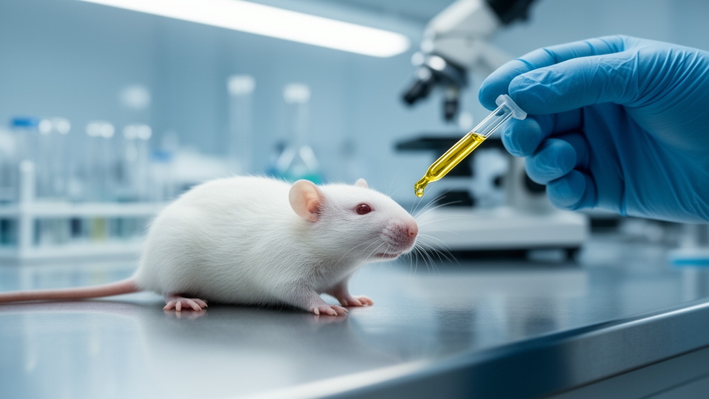
To test this, the investigators ran a controlled, prospective, randomized, interventional animal study involving 40 healthy adult male albino rats aged 3 to 4 months and weighing 170 to 200 grams. Animals with prior illness, dermatological abnormalities, or abnormal baseline liver enzymes were excluded. The rats were randomly assigned to four groups of ten each: a control group, an omega-3-only group, a heat-stroke group, and an omega-3-pretreated heat-stroke group. After 14 days of quarantine and acclimatization, treated animals received oral omega-3 at 400 milligrams per kilogram daily for 28 days, delivered through a dedicated oropharyngeal tube for each rat. Heat stroke was simulated by placing anesthetized rats in a controlled environmental chamber at 39.5 plus or minus 0.5 degrees Celsius for 180 minutes, with rectal core temperature continuously monitored. Animals were anesthetized with intraperitoneal sodium pentobarbital before heat exposure and euthanized according to ethical guidelines.
The biochemical core of the study focused on two familiar enzymes. Alanine aminotransferase, or ALT, sits predominantly in the hepatocyte cytoplasm and is relatively liver-specific, while aspartate aminotransferase, or AST, occurs in both cytoplasm and mitochondria and is abundant in liver, skeletal muscle, and erythrocytes. Both leak into the circulation when hepatocellular injury disrupts cell membranes, and both can continue rising after death as autolysis releases intracellular contents. Blood was drawn after euthanasia and samples were stored alongside the carcasses under the same experimental conditions, then analyzed at predetermined postmortem intervals of 0, 12, 24, 36, and 48 hours using a Beckman UniCel DxC800 Synchron system. Liver and skin samples were collected at intervals stretching from 0 to 168 hours, fixed in formalin, embedded in paraffin, sectioned at 5 to 6 micrometers, and stained with hematoxylin and eosin for blinded qualitative assessment by two independent observers, with disagreements resolved by a third.
The statistical results split cleanly down the middle. ALT and AST both increased progressively with advancing postmortem interval in every group, with p values below 0.001, confirming that these enzymes behave in a strongly time-dependent fashion after death. Heat-exposed rats showed significantly higher levels of both enzymes than control and omega-3-only animals. But the decisive finding came from a two-by-two factorial analysis treating heat stroke and omega-3 pretreatment as separate factors. For ALT, the Heat Stroke by Omega-3 interaction was highly significant, with an F value of 55.947 and p below 0.001, meaning the effect of omega-3 depended on whether the animal had experienced heat stroke.
AST told a different story. Although AST also climbed significantly over time and heat stroke produced a significant main effect, the Heat Stroke by Omega-3 interaction was not statistically significant (F = 0.765, p = 0.388). AST levels were numerically lower in the omega-3-pretreated heat-stroke group, but the differences from the heat-stroke group alone were not significant at any individual postmortem interval or overall (p = 0.162). The authors attribute this asymmetry partly to the biology of the two enzymes: ALT has a longer circulating half-life, roughly 40 to 60 hours in rats and comparable species, versus approximately 12 hours for AST, and AST is far more vulnerable to confounding from hemolysis and muscle injury because of its high erythrocyte content. ALT may therefore be the more informative marker for detecting acute and subacute hepatocellular injury in this setting.
The histological findings reinforced the biochemical picture, with an important caveat. In the heat-exposed groups, liver sections at the moment of death already showed distorted hepatic cords, severe congestion of blood sinusoids, cytoplasmic vacuolization, hydropic changes, focal fatty changes, and inflammatory infiltration. Autolysis then progressed faster in heat-exposed animals regardless of treatment: control and omega-3-only rats retained recognizable hepatic architecture up to about 72 hours postmortem, whereas heat-exposed groups showed disrupted organization and fragmented hepatic cords as early as 48 hours, and complete loss of recognizable architecture by 120 hours, at which point sampling stopped. In the omega-3-pretreated heat-stroke group, congestion, hemorrhage, and structural damage appeared less pronounced at corresponding intervals, with better-preserved hepatocyte contours and residual nuclei visible at time points where the untreated heat-stroke group had lost nearly all cellular detail.
Skin followed a parallel trajectory. Dermo-epidermal separation, dermal vacuolar degeneration, collagen fragmentation, and nuclear degeneration became increasingly evident beyond 72 hours, appearing earlier or more severely in the heat-stroke group, while the omega-3-pretreated heat-exposed group showed relatively better preservation at matching intervals. By 168 hours, both untreated groups showed complete epidermal-dermal separation, and by 120 hours both heat-exposed groups had reached severe autolysis, although tissue remained histologically identifiable as skin. Crucially, the authors repeatedly flag that these histological observations are qualitative. They explicitly state that the apparent preservation in treated animals does not independently demonstrate that omega-3 slowed postmortem autolysis, and may instead reflect reduced premortem heat-induced injury, meaning the tissue simply started the postmortem period in better condition. Interobserver agreement was also not formally quantified with a statistic such as Cohen’s kappa.
Beyond the treatment question, the study explored whether ALT and AST could serve as quantitative clocks for the postmortem interval. Pearson correlations showed strong positive associations between both enzymes and postmortem interval in all groups, and the team fitted linear and quadratic regression models predicting interval from enzyme levels, validated with nested leave-one-animal-out cross-validation to prevent data leakage. The omega-3-only group achieved the best internal performance, with a cross-validated root mean square error of 5.209 hours and a cross-validated R-squared of 0.906, followed by the heat-stroke group at 5.667 hours and 0.888. ALT was a significant predictor in all groups, while AST reached significance only in the two heat-exposed groups. The authors are careful to label these equations exploratory, internally validated models developed under controlled laboratory conditions that require external validation before any forensic application.
The broader forensic message is that antemortem nutritional and pathological states can leave fingerprints on postmortem measurements, complicating both interval estimation and perimortem injury assessment. The authors propose, as literature-supported hypotheses rather than demonstrated mechanisms, that omega-3’s reported antioxidant and anti-inflammatory activities, its suppression of NF-kappa-B signaling, and its incorporation into cell and mitochondrial membranes may explain the attenuated findings, but oxidative-stress markers, inflammatory mediators, and molecular pathways were not measured in this study. They also acknowledge the limits of the rat model, the small sample size, and the gap between controlled chamber conditions and the variability of real forensic casework. Larger studies with quantitative histological scoring, mechanistic biomarkers, independent validation cohorts, and human postmortem samples are needed before any of these findings could inform practice. For now, the study stands as a controlled demonstration that what a body experienced before death, and what it was fed beforehand, can measurably reshape the biochemical and histological evidence it leaves behind.
Subject of Research: The effect of omega-3 pretreatment on postmortem biochemical and histopathological changes in rats exposed to heat stroke
Article Title: Omega-3 pretreatment modulates postmortem biochemical and histopathological changes following heat stroke: an experimental study on adult male albino rats
Article References: Hassan, R. B., Abdel-Ghaffar, W. H., Badawy, A. H., Elrashedy, M. M., & Hadhoud, R. M. (2026). Omega-3 pretreatment modulates postmortem biochemical and histopathological changes following heat stroke: an experimental study on adult male albino rats. Egyptian Journal of Forensic Sciences, 16(1), Article 85. https://doi.org/10.1186/s41935-026-00584-z
Image Credits: AI Generated
DOI: 10.1186/s41935-026-00584-z
Keywords: forensic science, postmortem interval, heat stroke, omega-3, ALT, AST, autolysis, histopathology, rat model, liver injury, PMI estimation, hyperthermia
News Source: Cassandra Pierce. (October 29, 2026). Omega-3 Before Heat Stroke Shifts Postmortem Enzyme Patterns in Rats. Scienmag.



