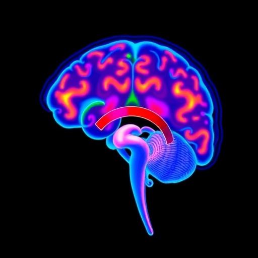
A new study from the University of Delaware suggests that the female brain may undergo subtle, measurable mechanical changes as estrogen levels rise and fall during the reproductive cycle. Using magnetic resonance elastography, or MRE, researchers detected hormone-sensitive changes in the hippocampus of rats, revealing that brain tissue may not simply respond to estrogen through biochemical signaling. It may also change in the way it physically deforms and moves under pressure. The findings, published in Brain Communications, offer an early glimpse into how reproductive hormones may reshape the brain’s structure and mechanical environment over time.
The hippocampus is best known for its role in memory and learning, but it is also one of the brain regions most responsive to hormonal signals. Estrogen receptors are widely distributed throughout the hippocampus, where estrogen can influence neuronal growth, synaptic plasticity and communication between brain cells. Synaptic plasticity refers to the ability of connections between neurons to strengthen, weaken or reorganize in response to experience. This process is considered essential for learning, memory formation and adaptation. The Delaware study adds a physical dimension to that biology by showing that the tissue surrounding these neural networks also appears to change across the rat oestrous cycle.
MRE is an advanced imaging method that extends the capabilities of conventional magnetic resonance imaging. During an MRE scan, gentle mechanical vibrations are transmitted through the body or brain. The MRI system then measures how those waves travel through tissue. Because softer and stiffer materials transmit mechanical waves differently, researchers can use the resulting patterns to calculate tissue properties such as stiffness, elasticity and viscoelasticity. These measurements provide information about how biological tissue responds to force. In the new study, the technique allowed researchers to examine the mechanical behavior of the hippocampus without removing tissue or disrupting the animals’ normal physiology.
The researchers found that hippocampal tissue mechanics varied in association with fluctuations in estrogen across the reproductive cycle. The result does not mean that estrogen acts as a simple switch that makes the hippocampus either soft or rigid. Brain tissue is a complex material made up of neurons, glial cells, blood vessels, extracellular matrix and fluid. Changes in any of these components could influence the way mechanical waves move through the region. Estrogen may affect several of them at once by altering neuronal structure, blood flow, cellular support systems or the organization of synapses. The MRE signal therefore offers a broad physical readout of biological changes occurring within the hippocampus.
“Across the reproductive cycle, the brain adapts, relying more heavily on different regions at different times,” said Katrina Milbocker, the study’s first author and a postdoctoral researcher in the laboratory of Curtis Johnson, an associate professor of biomedical engineering at the University of Delaware. Milbocker, who earned her doctorate in behavioral neuroscience at Delaware, said the work represents an initial test of whether MRE can track hormone-associated changes in the brains of rats before the approach is explored in humans. The study’s central contribution is thus methodological as well as biological: it demonstrates that a noninvasive imaging technique can detect cyclical mechanical variation in a hormone-sensitive brain structure.
The findings may be especially important because brain imaging is often used primarily to identify disease. MRE has been investigated in conditions such as tumors, neurodegenerative disorders and traumatic brain injury, where tissue mechanics can be altered. The Delaware researchers are pursuing a different application: using mechanical measurements to study healthy brain dynamics. “Imaging is often viewed as a tool for detecting disease, but it can also help us understand brain health dynamically,” Johnson said. “Our ultimate goal is to understand how brain mechanics change across life stages and what those changes can tell us about health and aging.” In this context, a change in tissue mechanics would not necessarily indicate damage. It could instead reflect normal adaptation, remodeling or altered cellular activity.
The study does not yet establish whether the observed mechanical changes improve or impair memory and learning. That question will require experiments that combine MRE measurements with behavioral testing. Researchers could compare tissue mechanics with performance on tasks that assess spatial memory, learning speed or cognitive flexibility at different stages of the reproductive cycle. Such studies may reveal whether specific mechanical states correspond to changes in hippocampal function, or whether the imaging signal reflects biological remodeling that occurs without a measurable effect on behavior. Establishing that connection is essential before MRE can be considered a tool for evaluating hormone-related cognitive changes.
The team is now extending its work to rat models of menopause, when ovarian estrogen production falls substantially. Menopause is not an abrupt biological event for every individual; the transition can unfold over years and involve changing hormone levels, sleep disruption, mood symptoms and cognitive complaints. Many people report experiences commonly described as “brain fog,” although the causes and severity vary widely. Johnson said the researchers suspect that estrogen depletion could produce a mechanical “stuck state” in the brain, in which the hippocampus fails to adapt normally after hormone levels decline. This remains a hypothesis, however, and the planned experiments are intended to determine whether mechanical changes accompany or contribute to cognitive disruption.
The researchers also hope to move from animal studies toward human applications. Translating MRE findings across species will require careful work because the human brain is larger, more structurally complex and subject to considerable variation in hormone history, age, health and medication use. Human studies would need to examine whether similar hippocampal patterns appear across the menstrual cycle, during perimenopause and after menopause. They could also investigate how hormone therapy, aging or neurological disease affects the measurements. The University of Delaware has an unusual logistical advantage for this effort: its human and animal imaging facilities are located in the same building, allowing researchers to coordinate protocols and compare results more efficiently.
For MRE to become useful in routine clinical research or medical care, the scanning process will also need to be fast, reliable and easy to integrate with standard MRI examinations. The Delaware team hopes to develop an approach that adds less than a minute to a conventional scan. If that goal can be achieved, mechanical measurements could eventually be collected alongside images of brain anatomy and activity. Such information might help researchers follow how the brain responds to major hormonal transitions, including menopause, and identify patterns associated with healthy aging or cognitive vulnerability. For now, the study provides an early but striking message: the hormonal life of the brain may be visible not only in its chemistry and circuitry, but also in the physical behavior of its tissue.
Subject of Research: Estrogen-related changes in hippocampal tissue mechanics across the rat oestrous cycle, measured using magnetic resonance elastography.
Article Title: Hippocampal tissue mechanics are sensitive to fluctuations in oestrogen across the rat oestrous cycle
Web References: https://academic.oup.com/braincomms/advance-article/doi/10.1093/braincomms/fcag228/8711977; https://engr.udel.edu/news/2026/08/tracking-estrogens-effects-on-the-brain/
References: Brain Communications, DOI: 10.1093/braincomms/fcag228
Keywords
Estrogen, hippocampus, magnetic resonance elastography, brain mechanics, neuroscience, reproductive cycle, menopause, memory, cognitive health, biomedical engineering
Tags: estrogen effects on hippocampal structureestrogen receptors and neural connectivityhippocampus response to hormonal fluctuationshormone-driven brain changeshormone-sensitive brain plasticitymagnetic resonance elastography in neurosciencemechanical properties of brain tissueneuroimaging techniques for hormonal effectsnovel imaging methods in brain researchphysical deformation of brain tissuereproductive cycle and brain mechanicsreproductive hormones and memory formation