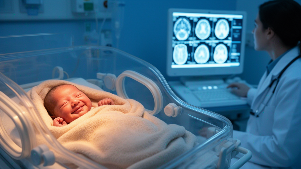
For parents of a baby born weeks or months too early, few moments in the neonatal intensive care unit carry as much emotional weight as the cranial ultrasound. The painless scan, performed through the soft fontanelle at the top of the skull, offers the first glimpse of the infant’s developing brain, and an abnormal finding can cast a long shadow over the weeks that follow. Yet the reassurance offered by a normal scan has always been difficult to quantify. How much comfort should a family actually take from a clean first image, and does that reassurance grow stronger each time the scan comes back normal? A large new study from Spain now provides some of the most detailed answers to date, and its findings are both encouraging and carefully nuanced.
Researchers at Hospital Universitario 12 de Octubre in Madrid analysed data from a prospectively maintained neonatal and neurodevelopmental follow-up registry spanning nearly three decades, from 1991 to 2020. Their cohort included 3,081 live-born infants who arrived early in the world, defined as those born before 32 weeks of gestation or weighing 1,500 grams or less at birth. Of these, 2,841 had complete data from their first cranial ultrasound, and the vast majority went on to receive serial scans throughout their neonatal course. The study, published as an open-access research article in BMC Pediatrics, examined whether a pattern the investigators call persistent normality, meaning normal findings maintained across the entire serial ultrasound pathway, could serve as a marker of a low-risk early developmental trajectory.
The technical architecture of the surveillance protocol is central to understanding the results. Cranial ultrasound was performed in four predefined windows during the neonatal admission, beginning with a scan within 48 hours of birth, followed by examinations around day 7 and day 28, with a further scan later in the hospital stay. At each time point, radiologists and clinicians recorded the presence or absence of three categories of findings: intraventricular haemorrhage, which is bleeding into the fluid-filled spaces of the brain; ventriculomegaly, an abnormal enlargement of those spaces that can signal rising pressure or tissue loss; and parenchymal lesions, which involve damage to the brain tissue itself. Because injury patterns in preterm brains evolve over days and weeks, a single early scan can miss problems that emerge later, which is precisely why serial imaging has become standard practice in neonatal units worldwide.
The headline numbers are striking. Among the 1,881 infants whose first ultrasound was completely normal, representing 66.2 percent of those with first-scan data, 97.4 percent went on to have subsequent follow-up imaging. Within that group, 70.7 percent maintained a completely normal trajectory across all their serial scans. That probability climbed steadily with each additional normal result: after two consecutive normal scans, 80.8 percent remained normal throughout, and after three, the figure rose to 91.7 percent. In other words, every clean scan compounds the statistical likelihood that the brain will remain free of the injuries that ultrasound can detect, a pattern that will resonate with clinicians who have long suspected but never fully quantified this cumulative reassurance.
Gestational age at birth proved to be a powerful modifier of these probabilities. Among infants born at 24 to 25 weeks of gestation, the most extreme end of prematurity, only 42.3 percent maintained normality after an initial normal scan. By contrast, among those born at 30 weeks or later, the figure reached 82.2 percent. This gradient makes biological sense: the earlier the brain is delivered into the extrauterine environment, the more vulnerable its immature white matter and fragile germinal matrix are to haemorrhage and injury, even in babies whose first images look pristine. For counselling purposes, this means the same normal first scan carries very different prognostic weight depending on how early the child was born.
The most consequential findings concern long-term neurodevelopment. The researchers adjusted their analyses for gestational age and birth epoch, allowing them to isolate the association between ultrasound trajectory and outcomes. Children whose serial cranial ultrasounds remained persistently normal had dramatically lower rates of cerebral palsy at two years of age: 0.7 percent, compared with 11.9 percent among those without persistent normality. After adjustment, this translated to an odds ratio of 0.07, meaning persistently normal infants had roughly fourteen-fold lower odds of cerebral palsy. Rates of late or absent independent walking were also reduced, at 4.0 percent versus 8.6 percent, and the need for sustained developmental or educational support during early childhood was less than half as frequent, at 10.0 percent versus 21.9 percent. Around the transition from preschool to school age, cognitive sequelae appeared in 6.8 percent of the persistently normal group versus 15.2 percent of the comparison group.
Equally informative was what happened to infants whose first scan was normal but who later developed abnormalities. Across the broader cohort, these children showed outcome frequencies close to those whose first scan was abnormal from the start. This observation carries an important clinical message: a normal early scan does not confer lasting protection if later imaging reveals injury, and the developmental risk tracks with the eventual brain findings rather than with the initial reassuring image. It underscores why the serial nature of the surveillance matters, and why clinicians should resist anchoring their prognostic expectations on the first scan alone.
The study is equally candid about the limits of ultrasound-based reassurance. Persistent normality did not clearly discriminate several important outcomes, including documented or suspected autism spectrum disorder, hyperactive or inattentive behavioural concerns noted at preschool age, learning difficulties after statistical adjustment, or sensory impairment. This is not surprising to developmental neuroscientists. Cranial ultrasound excels at detecting gross structural lesions such as haemorrhage and ventricular dilation, but many of the subtle disruptions to connectivity and cortical development that underlie social-communication difficulties and behavioural vulnerabilities are simply invisible to the modality. A structurally normal brain on ultrasound can still carry functional differences that emerge only as the child grows and faces increasingly complex cognitive and social demands.
The authors frame their conclusions as support for risk-stratified follow-up rather than for discharge from surveillance. Persistent normality identifies a low-risk trajectory, they emphasise, but a low-risk trajectory is not a risk-free one. In practical terms, the findings could help neonatal follow-up programmes calibrate the intensity of monitoring: families of children with persistently normal imaging may be counselled that major motor impairment is unlikely, while still being told that behavioural, social-communication, language and educational vulnerabilities can appear regardless of scan results. Conversely, infants whose normality breaks down on serial imaging warrant closer developmental surveillance, since their outcome profile resembles that of children with abnormal scans from the outset.
For the families whose children populate this three-decade registry, the study distils a message that is both comforting and sobering. Each normal ultrasound genuinely shifts the odds toward a healthier developmental path, particularly for major motor outcomes and the need for early-childhood support, and the reassurance grows with every consecutive clean scan. But the developing brain keeps some of its secrets hidden from sound waves, and no imaging finding, however reassuring, replaces the careful, ongoing observation of how a child moves, communicates, learns and connects with the world. The Madrid team’s work gives clinicians a quantified foundation for that delicate balance between hope and vigilance, and it reminds parents that a normal scan is a strong signal, never a final verdict.
Subject of Research: Serial cranial ultrasound normality as a predictor of neurodevelopmental outcomes in preterm infants
Article Title: Persistent normality on serial cranial ultrasound as a marker of early developmental trajectory in preterm infants: a single-centre retrospective cohort study
Article References: Núñez-Enamorado, N., Camacho-Salas, A., López-Maestro, M., Gallego-Herrero, M. C., Zamora-Crespo, B., Vila-Bedmar, S., Vázquez-Román, S., de Aragón, A. M., Pallás-Alonso, C. R., & Moral-Pumarega, M. T. (2026). Persistent normality on serial cranial ultrasound as a marker of early developmental trajectory in preterm infants: a single-centre retrospective cohort study. BMC Pediatrics. https://doi.org/10.1186/s12887-026-07692-8
Image Credits: AI Generated
DOI: 10.1186/s12887-026-07692-8
Keywords: preterm infants, cranial ultrasound, neurodevelopment, cerebral palsy, developmental trajectory, neonatal neuroimaging, cognitive sequelae, educational support, early childhood, follow-up, ventriculomegaly, intraventricular haemorrhage
News Source: Cassandra Pierce. (October 15, 2026). Normal Brain Scans Over Time Point to Brighter Outlook for Preterm Babies. Scienmag.