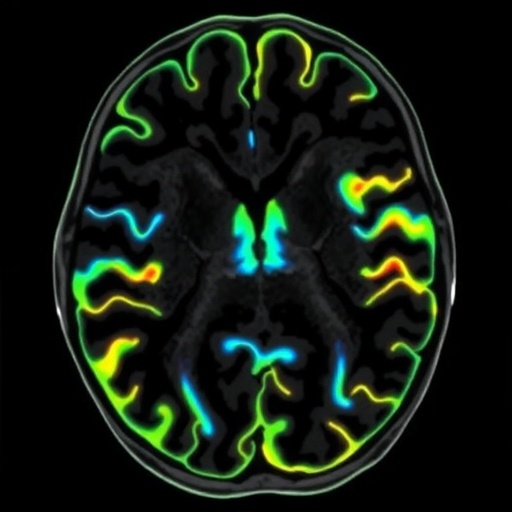
A little-known anatomical feature surrounding the brain is moving into the spotlight as researchers investigate how it can be studied without surgery, injections or other invasive procedures. In a study published in Nature Communications, N.E. Fultz, G. Ringstad, M. Debiasi and colleagues examine the perivascular subarachnoid spaces—tiny fluid-containing compartments located around blood vessels as they pass through the brain’s subarachnoid space. Their work focuses on how these structures can be characterized non-invasively, potentially opening a new window onto the relationship between blood vessels, cerebrospinal fluid and neurological disease.
The subarachnoid space is best known as the layer between two protective membranes surrounding the brain and spinal cord. It contains cerebrospinal fluid, or CSF, which cushions the central nervous system and participates in the movement of nutrients, signaling molecules and waste products. Within this environment, arteries and veins travel along the brain’s surface and branch into deeper tissue. The spaces surrounding some of these vessels form specialized anatomical corridors that may influence fluid movement and communication between the brain’s vascular and fluid systems.
Researchers have long recognized that fluid does not circulate through the brain in a simple, open plumbing network. Instead, CSF moves through interconnected compartments, while blood vessels create boundaries and pathways that can shape local flow. Perivascular spaces are particularly important because they lie at the interface of vascular pulsation, tissue structure and fluid transport. Changes in their size, shape or visibility may reflect alterations in pressure, inflammation, vascular function or the clearance of metabolic waste.
The phrase “perivascular subarachnoid spaces” refers specifically to spaces associated with vessels in the subarachnoid compartment, rather than the more commonly discussed perivascular spaces located within the brain’s white matter and deep gray matter. Distinguishing these regions is technically important. Similar-looking spaces can arise in different anatomical locations and may have different biological meanings. A method that can reliably identify and characterize them could help researchers separate normal anatomical variation from changes linked to disease.
The study’s central significance lies in its non-invasive approach. Instead of relying on tissue removal or direct surgical access, non-invasive characterization generally uses advanced medical imaging and quantitative analysis to extract anatomical and physiological information from living participants. Imaging can reveal the geometry of fluid spaces, their relationship to nearby vessels and, in some circumstances, indirect signs of fluid movement. Such measurements are especially valuable for studying structures that are too small, delicate or inaccessible to investigate directly in routine clinical practice.
This type of research could also help clarify how the brain’s waste-clearance systems operate. The glymphatic system, a proposed network involving CSF and interstitial fluid, has attracted intense interest because it may help transport metabolic by-products away from neural tissue. Perivascular pathways are thought to be involved in this process, although their exact roles, direction of flow and relationship to other fluid compartments remain active areas of investigation. Better imaging of the spaces around surface vessels may provide data needed to test competing explanations rather than relying solely on theoretical models.
The potential medical relevance is broad. Disturbances in cerebrospinal-fluid dynamics and vascular function appear in conditions ranging from hydrocephalus and stroke to small-vessel disease, traumatic brain injury and neurodegenerative disorders. Enlarged or altered perivascular spaces have also been reported in association with aging and several brain diseases. However, an imaging finding is not automatically a diagnostic marker. Researchers must determine how much variation is normal, whether measurements are reproducible between scanners and observers, and whether changes in these spaces predict symptoms or clinical outcomes.
A reliable non-invasive method could eventually make it easier to compare the brain’s fluid compartments across individuals and over time. Longitudinal imaging might allow scientists to observe whether perivascular structures change with age, sleep, blood-pressure control or disease progression. It could also support studies of therapies designed to influence vascular pulsatility, CSF circulation or waste clearance. For now, the value of the work is primarily methodological: before a biological structure can become a biomarker, investigators need a consistent way to see and measure it.
The findings arrive at a moment when brain imaging is becoming increasingly quantitative. Modern scanners can generate high-resolution anatomical maps, while computational techniques can identify subtle structures and calculate their spatial relationships. Yet greater technical power also creates new challenges, including the risk of confusing imaging artifacts with anatomy and the danger of attaching biological meaning to patterns that have not been independently validated. Studies such as this one are therefore important not only because they highlight a hidden component of brain organization, but also because they help establish the measurement standards required for future research.
Perivascular subarachnoid spaces may sound like a specialized anatomical detail, but they sit at a potentially crucial crossroads linking blood vessels, cerebrospinal fluid and the brain’s protective membranes. By pursuing a non-invasive way to study them, Fultz, Ringstad, Debiasi and their colleagues are contributing to a broader effort to make the brain’s fluid circulation visible in living people. The approach does not yet transform these spaces into a clinical test, but it could provide researchers with a new tool for investigating how the brain maintains its internal environment—and what happens when that finely balanced system begins to fail.
Subject of Research: Non-invasive characterization of perivascular subarachnoid spaces in the human brain.
Article Title: Non-invasive characterization of perivascular subarachnoid spaces
Article References: Fultz, N.E., Ringstad, G., Debiasi, M. et al. “Non-invasive characterization of perivascular subarachnoid spaces.” Nature Communications (2026). https://doi.org/10.1038/s41467-026-76306-9
Image Credits: AI Generated
DOI: 10.1038/s41467-026-76306-9
Keywords: perivascular subarachnoid spaces, cerebrospinal fluid, brain imaging, neuroanatomy, vascular biology, glymphatic system, non-invasive research, neurological disease
Tags: brain fluid dynamicsbrain vascular anatomycerebrospinal fluid circulationcerebrospinal fluid pathwaysMRI-based brain imagingneuroimaging techniquesneurological disease biomarkersneurovascular couplingNoninvasive brain imagingperivascular space characterizationperivascular spacessubarachnoid space