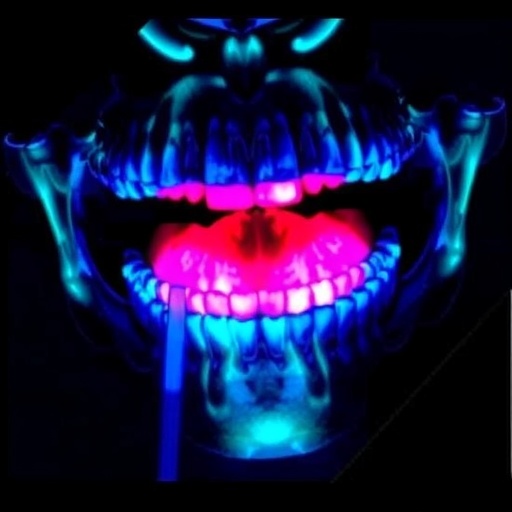
In a groundbreaking advancement for oncological surgery, researchers have unveiled a novel near-infrared fluorescence (NIRF) imaging-guided technique employing a cRGD-ZW800 conjugate to significantly enhance the precision of surgical resections in oral cancer. This innovative approach, recently rigorously tested in a phase I/II feasibility trial, offers a glimpse into the future of surgical oncology, where real-time, high-resolution visualization of tumor margins could become the gold standard for improving patient outcomes and reducing recurrence rates.
The challenge of achieving clear surgical margins in oral cancer has long plagued surgeons, as the anatomical complexity and the infiltrative nature of tumors make it exceedingly difficult to visually demarcate malignant from healthy tissue. Traditional methods rely heavily on tactile feedback and histopathological examination post-surgery, leading to high reoperation rates and suboptimal prognosis. The integration of molecular imaging directly into the surgical workflow stands as a transformative solution, enabling surgeons to delineate tumor boundaries with unprecedented accuracy.
Central to this technique is the use of cRGD-ZW800, a molecular probe that selectively binds to integrin receptors, prominently expressed on the surface of cancerous cells and their neovasculature. Chlorotoxin-based and RGD peptide derivatives have previously been explored for tumor targeting, but the cRGD modification here provides enhanced affinity and specificity toward αvβ3 integrin, a critical mediator of angiogenesis and tumor progression. The conjugation to ZW800, a near-infrared fluorescent dye characterized by high quantum yield and excellent photostability, allows for deep tissue penetration and minimal autofluorescence, pivotal qualities for effective in vivo imaging.
The phase I/II trial encompassed a cohort of patients diagnosed with oral squamous cell carcinoma, undergoing surgical resection. Prior to surgery, patients received intravenous administration of the cRGD-ZW800 probe, allowing the agent to accumulate within tumor tissues. During surgery, a dedicated NIRF imaging system was employed to visualize the fluorescent signal emitted by the probe, highlighting cancerous regions against a low-fluorescence background. Surgeons utilized this real-time feedback to guide resection, aiming to capture all malignant tissue while preserving healthy structures.
Quantitative assessment of surgical margins demonstrated a marked improvement when guided by NIRF imaging, with a significantly higher rate of negative margins compared to historical controls. Notably, this approach enabled the identification of subclinical satellite lesions and infiltrative tumor extensions that would have otherwise escaped detection via conventional white-light visualization. The ability to detect microscopic disease at the margins intraoperatively minimizes the likelihood of residual tumor and the need for adjuvant therapies such as radiotherapy or chemotherapy.
In addition to technical performance, safety and pharmacokinetics of cRGD-ZW800 were scrutinized. The probe exhibited a favorable safety profile, with no serious adverse events attributable to the contrast agent reported. Rapid clearance from non-target tissues and retention within neoplastic areas ensured an optimal tumor-to-background ratio during the critical surgical timeframe. Such properties underscore the clinical translatability of this imaging probe for widespread adoption.
This trial also addressed potential limitations inherent in fluorescence-guided surgery, including signal attenuation due to tissue depth and light scattering. The near-infrared window utilized here strikes a balance between penetration depth and resolution, enabling visualization of tumors located several millimeters beneath the tissue surface. Coupled with advanced imaging hardware, the system facilitated intuitive integration into the surgical workflow without prolonging operative times.
The implications of this study extend beyond oral cancer, as the molecular targeting strategy and imaging technology hold promise for other solid tumors characterized by integrin overexpression. By refining the accuracy of surgical excision, the approach could drastically reduce local recurrence rates, improve survival, and enhance quality of life for cancer patients. Moreover, the precision offered may democratize complex oncological resections, potentially allowing surgeons in diverse settings to achieve superior outcomes.
Future directions include optimization of probe dosing and administration timing, exploration of multimodal imaging combinations, and integration with robotic-assisted surgical systems. Enhancements in real-time image processing and display interfaces will further empower surgeons to make informed intraoperative decisions. Large-scale randomized trials are anticipated to validate these findings and solidify regulatory approval pathways.
On a mechanistic level, the success of cRGD-ZW800 highlights the therapeutic potential of targeting the tumor microenvironment, particularly angiogenic pathways. Integrin αvβ3 not only facilitates cell adhesion and migration but also modulates signaling networks pivotal to tumor survival. By visualizing this biomarker, surgeons gain insight into tumor biology that transcends morphology alone, ushering in an era of functional surgery.
In conclusion, the advent of near-infrared fluorescence imaging-guided surgery using cRGD-ZW800 represents a paradigm shift in oncological interventions for oral cancer. This technique merges molecular targeting with cutting-edge optical imaging, enabling surgeons to surpass the limitations of conventional surgical visualization. The phase I/II feasibility trial serves as a crucial milestone, demonstrating feasibility, safety, and clinical benefit of this approach. As technological refinements and broader clinical validation proceed, this innovation is poised to transform the landscape of cancer surgery fundamentally.
This pioneering work exemplifies the convergence of molecular biology, photonics, and surgical innovation, illustrating how interdisciplinary collaboration can yield transformative healthcare solutions. For patients battling oral cancer, where recurrence and morbidity remain daunting challenges, fluorescence-guided surgery offers renewed hope. This breakthrough sets a precedent for harnessing the power of targeted imaging to deliver precision medicine directly into the operating room.
As the field of fluorescence-guided oncology evolves, the integration of targeted probes like cRGD-ZW800 with artificial intelligence-driven image analysis and decision support may further elevate surgical precision. The future envisions a fully integrated surgical environment where multimodal imaging, real-time analytics, and robotic assistance converge seamlessly, ultimately improving outcomes across a spectrum of malignancies.
This research ushers in a promising new chapter in cancer surgery, with near-infrared fluorescence imaging emerging as a critical tool in the surgeon’s arsenal. The meticulous work and clinical insight driving cRGD-ZW800 development pave the way for broader application, signaling a paradigm shift that could redefine standards of care in oncology worldwide.
Subject of Research: Near-infrared fluorescence imaging for improved surgical resection margins in oral cancer using a targeted molecular probe (cRGD-ZW800)
Article Title: Near-infrared fluorescence imaging-guided surgery using cRGD-ZW800 to improve surgical resection margins in oral cancer: a phase I/II feasibility trial
Article References:
Zweedijk, B.E., Lauwerends, L.J., Galema, H.A. et al. Near-infrared fluorescence imaging-guided surgery using cRGD-ZW800 to improve surgical resection margins in oral cancer: a phase I/II feasibility trial. Nat Commun (2026). https://doi.org/10.1038/s41467-026-73554-7
Image Credits: AI Generated
Tags: cRGD-ZW800 molecular probefluorescence-guided tumor resectionintegrin-targeted imagingmolecular imaging in cancernear-infrared fluorescence imagingoncological surgery innovationoral cancer surgeryphase I/II clinical trialreal-time surgical guidancereducing oral cancer recurrencesurgical margin precisiontumor margin visualization

