• HOME
  • NEWS
  • EXPLORE
    • CAREER
      • Companies
      • Jobs
    • EVENTS
    • iGEM
      • News
      • Team
    • PHOTOS
    • VIDEO
    • WIKI
  • BLOG
  • COMMUNITY
    • FACEBOOK
    • INSTAGRAM
    • TWITTER
Sunday, October 4, 2026
BIOENGINEER.ORG
No Result
View All Result
  • Login
  • HOME
  • NEWS
  • EXPLORE
    • CAREER
      • Companies
      • Jobs
        • Lecturer
        • PhD Studentship
        • Postdoc
        • Research Assistant
    • EVENTS
    • iGEM
      • News
      • Team
    • PHOTOS
    • VIDEO
    • WIKI
  • BLOG
  • COMMUNITY
    • FACEBOOK
    • INSTAGRAM
    • TWITTER
No Result
View All Result
Bioengineer.org
No Result
View All Result
Home NEWS Science News

New software automates brain imaging

Bioengineer by Bioengineer
February 6, 2017
in Science News
Reading Time: 2 mins read
0
Share on FacebookShare on TwitterShare on LinkedinShare on RedditShare on Telegram
IMAGE

Credit: Max Planck Florida Institute for Neuroscience

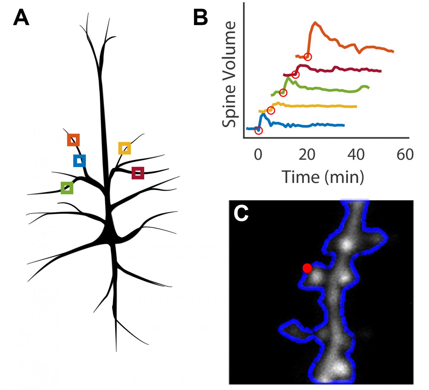
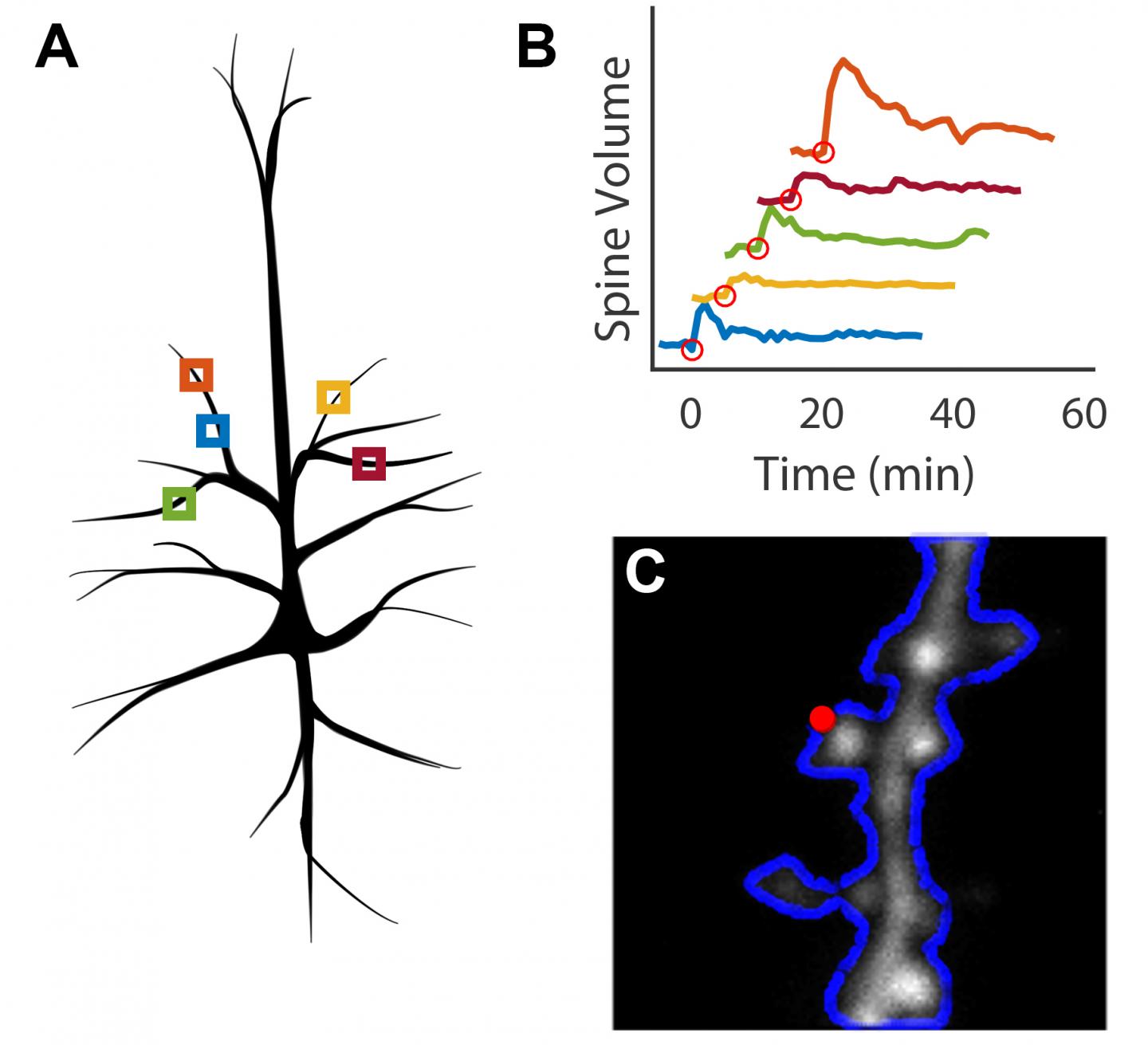
When humans and animals learn and form memories, the physical structures of their brain cells change. Specifically, small protrusions called dendritic spines, which receive signals from other neurons, can grow and change shape indefinitely in response to stimulation. Scientists at Max Planck Florida Institute for Neuroscience (MPFI) have observed this process, known as long-term structural plasticity, in individual spines, but doing so requires substantial time and effort. A new technique, developed by MPFI researchers, automates the process to make observing and quantifying this growth far more efficient. The open-source method is available to any scientist hoping to image plasticity as it happens in dendritic spines using Scanimage. The work was published in January 2016 in the Public Library of Science journal, PLOS ONE.

Scientists working in Ryohei Yasuda's laboratory at MPFI are working to understand how proteins facilitate the plasticity of dendritic spines, the biological basis of learning and memory. They use 2-photon microscopy, an advanced technique for live-cell imaging, and glutamate uncaging, a technique that can induce plasticity in individual spines of interest using light. This is a meticulous process, wherein a scientist must continually focus the microscope on a single dendritic spine over an extended period, often an hour or longer. Michael Smirnov, Ph.D., Post-doctoral researcher at MPFI, developed a software that allows the computer to automatically track, image, and stimulate up to five dendritic spines at a time. "We can collect the data and figure out the proteins responsible much quicker with this [program] because we can run much more robust experiments," said Smirnov. In addition to increasing productivity, the ability to stimulate and image multiple spines in parallel greatly decreases the cost of running these experiments.

The software is a MATLAB-based module built for Scanimage, a program already commonly used in life science laboratories. It includes an electrically tunable lens in combination with a drift correction algorithm. These aspects allow the program to identify and correct for sample movement to ensure that the microscope is consistently focused on the spines of interest throughout the duration of the experiment. The interface provides an inexpensive method for automating experiments that observe up to five dendritic spines at a time, as opposed to a single spine using existing methods.

In contrast to previous open-sourced focusing programs, this one implements a highly capable and customizable focus and drift correction system to ensure that it can be used for a variety of biological applications. "The paper explains further modifications to make the process automated," said Smirnov. "It shares the open source code, so essentially, other people from other institutes can easily pick this up and use it for themselves."

###

About Max Planck Florida Institute for Neuroscience

The Max Planck Florida Institute for Neuroscience (Jupiter, Florida, USA) specializes in the development and application of novel technologies for probing the structure, function, and development of neural circuits. It is the first research institute of the Max Planck Society in the United States.

Media Contact

Jennifer Gutierrez
[email protected]
@mpfneuro

http://www.maxplanckflorida.org/

############

Story Source: Materials provided by Scienmag

Share12Tweet8Share2ShareShareShare2

Related Posts

Hydrogen Sulfide's Hidden Hand: How a Tiny Gas Molecule Rewires Plant Proteins

Hydrogen Sulfide’s Hidden Hand: How a Tiny Gas Molecule Rewires Plant Proteins

October 4, 2026
Junk Food May Age the Ovary Before the Egg Reserve Runs Dry

Junk Food May Age the Ovary Before the Egg Reserve Runs Dry

October 4, 2026

Flickering LEDs Boost Photosynthesis When Pulses Stay Under a Millisecond

October 4, 2026

Aspirin May Slow the Aging Clocks of Colon Cells, Study Suggests

October 4, 2026
Please login to join discussion

POPULAR NEWS

  • Alloys That Shrink Their Own Grains: New PIX Mechanism Refines Metals With Heat Alone

    Alloys That Shrink Their Own Grains: New PIX Mechanism Refines Metals With Heat Alone

    29 shares
    Share 12 Tweet 7
  • Endurance Exercise Reshapes the Liver in Males and Females Through Distinct Molecular Routes

    29 shares
    Share 12 Tweet 7
  • Single Transcription Factor PU.1 Rapidly Converts Fibroblasts into Macrophage-Lineage Cells

    29 shares
    Share 12 Tweet 7
  • New Scale Measures How Ready Nurse Educators Really Are for the AI Era

    29 shares
    Share 12 Tweet 7

About

We bring you the latest biotechnology news from best research centers and universities around the world. Check our website.

Follow us

Recent News

Alloys That Shrink Their Own Grains: New PIX Mechanism Refines Metals With Heat Alone

Endurance Exercise Reshapes the Liver in Males and Females Through Distinct Molecular Routes

Single Transcription Factor PU.1 Rapidly Converts Fibroblasts into Macrophage-Lineage Cells

Subscribe to Blog via Email

Success! An email was just sent to confirm your subscription. Please find the email now and click 'Confirm' to start subscribing.

Join 85 other subscribers
  • Contact Us

Bioengineer.org © Copyright 2023 All Rights Reserved.

Welcome Back!

Login to your account below

Forgotten Password?

Retrieve your password

Please enter your username or email address to reset your password.

Log In
No Result
View All Result
  • Homepages
    • Home Page 1
    • Home Page 2
  • News
  • National
  • Business
  • Health
  • Lifestyle
  • Science

Bioengineer.org © Copyright 2023 All Rights Reserved.