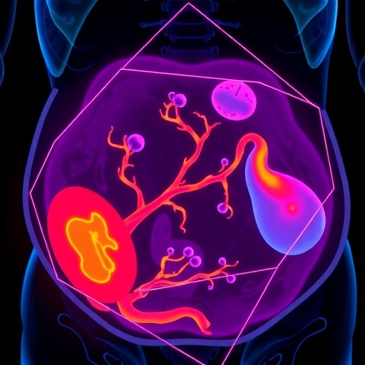
In a groundbreaking advancement for maternal-fetal medicine, researchers at Baylor College of Medicine have unveiled a novel artificial intelligence (AI) model capable of accurately detecting placenta accreta spectrum (PAS) before delivery. PAS, a perilous pregnancy complication marked by the abnormal adherence of the placenta to the uterine wall, has long posed diagnostic challenges, contributing significantly to maternal mortality and morbidity globally. This pioneering AI-driven diagnostic tool, presented at the 2026 Society for Maternal-Fetal Medicine (SMFM) Pregnancy Meeting™, represents a transformative step toward early identification and intervention, potentially revolutionizing prenatal care for high-risk pregnancies.
Placenta accreta spectrum encompasses a range of conditions wherein the placenta invades the uterine wall to varying degrees, often linked to prior uterine surgical history such as cesarean deliveries. The increasing prevalence of PAS in the United States, partly driven by higher cesarean rates, has intensified clinical urgency to develop reliable screening methods. Traditional approaches rely heavily on risk factor assessment and sonographic evaluation, yet these methods suffer from significant limitations, including operator dependency and the potential for inconclusive or misleading ultrasound findings. Consequently, nearly half of PAS cases remain undiagnosed until delivery, by which time catastrophic hemorrhage and other life-threatening complications may ensue.
Facing these diagnostic challenges, the Baylor research team employed an innovative AI algorithm designed to analyze two-dimensional (2D) obstetric ultrasound images with unprecedented accuracy. The retrospective study examined ultrasound images obtained from 113 pregnant patients identified as high risk for PAS, all of whom delivered at Texas Children’s Hospital between 2018 and 2025. On average, the ultrasounds were conducted around 31 weeks of gestation, a critical window in prenatal monitoring. The AI model’s analytical framework integrates deep learning techniques to discern subtle morphological patterns indicative of placental invasion that might elude human observers.
Results from the study were striking: the AI model successfully detected every confirmed case of PAS among the cohort, achieving perfect sensitivity. While the model generated two false positive cases, it notably avoided any false negatives, underscoring its potential as a highly reliable screening adjunct. This level of diagnostic precision could enable obstetricians to anticipate and prepare for complicated deliveries more effectively, thereby mitigating the risk of severe maternal hemorrhage, multi-organ failure, and mortality associated with undiagnosed PAS.
The methodological prowess of the AI model lies in its capacity to process complex imaging data beyond conventional visual analysis. By harnessing vast arrays of pixel-level information and training on annotated datasets, the algorithm learns to differentiate between normal placental attachment and pathological adherence. This transcends the variability inherent in human interpretation, offering a standardized and reproducible diagnostic tool. Furthermore, incorporating AI into obstetric ultrasound workflow could democratize expertise, providing critical decision support in settings where subspecialty maternal-fetal medicine consultation is scarce.
Dr. Alexandra L. Hammerquist, a maternal-fetal medicine fellow and lead researcher on the project, emphasized the clinical impact of this innovation. She stated, “Our team is very excited about the potential clinical implications of this model for accurate and timely diagnosis of PAS. We are hopeful that its use as a screening tool will help decrease PAS-related maternal morbidity and mortality.” The promise of AI-assisted diagnosis extends beyond simply detecting PAS; it opens the door to personalized prenatal care pathways, enabling tailored surveillance intensity and delivery planning.
The study’s retrospective design leveraged a comprehensive dataset amassed over nearly seven years, reflecting real-world clinical heterogeneity. The inclusion criteria focused on pregnancies deemed high risk due to clinical or obstetric history, making the findings particularly relevant for targeted screening strategies. The choice of 2D ultrasound, rather than more advanced imaging modalities, enhances the applicability of this AI tool, as 2D ultrasound remains the standard imaging technique worldwide due to its accessibility and cost-effectiveness.
While the occurrence of two false positives indicates room for refinement, the absence of false negatives is a critical attribute from a clinical safety perspective, ensuring that cases of PAS do not go undetected. Future prospective studies will be essential to validate these promising results, assess the AI model’s performance across diverse populations, and evaluate integration into clinical workflows. Additionally, exploring real-time application during ultrasound acquisition represents a thrilling frontier, potentially enabling instantaneous diagnostic support.
The implications of this research extend beyond PAS, illustrating the broader potential for AI to transform prenatal diagnostics. By augmenting human expertise with machine learning, clinicians can uncover subtle pathological features invisible to the naked eye, enhancing early detection of a spectrum of pregnancy complications. This study advances the paradigm toward precision obstetrics, where data-driven insights drive optimized therapeutic decisions, ultimately improving outcomes for both mothers and their babies.
This pioneering AI model has garnered significant attention ahead of its detailed presentation in oral abstract #39 titled “AI-based ultrasound screening for early, accurate identification of placenta accreta spectrum,” scheduled for publication in the February 2026 issue of Pregnancy, the official peer-reviewed journal of the Society for Maternal-Fetal Medicine. The research symbolizes a beacon of hope in maternal health, aiming to reduce the devastating sequelae of undiagnosed PAS and set a new standard for diagnostic accuracy in high-risk obstetrics.
With the rising global cesarean delivery rates and the concomitant surge in PAS incidence, timely and accurate diagnosis becomes paramount. The integration of AI into obstetric practice, as exemplified by this study, highlights a future wherein technology not only supports but also fundamentally reshapes clinical paradigms. As the medical community continues to grapple with complex pregnancy complications, innovations like the Baylor College model offer a compelling vision of safer pregnancies through enhanced early detection.
The Society for Maternal-Fetal Medicine, representing over 6,500 specialists devoted to managing high-risk pregnancies, underscores the critical need for such advancements by championing research, education, and advocacy in this field. This AI-based screening breakthrough thus sits at the intersection of clinical necessity and technological possibility, poised to deliver meaningful benefits for maternal and fetal health worldwide.
Subject of Research: People
Article Title: AI-based ultrasound screening for early, accurate identification of placenta accreta spectrum
News Publication Date: February 12, 2026
Web References: https://smfm2026.eventscribe.net/
References: Oral abstract #39 presented at the 2026 Pregnancy Meeting™, February 2026 issue of Pregnancy
Image Credits: Not provided
Keywords: Placenta accreta spectrum, AI model, prenatal diagnosis, obstetric ultrasound, maternal-fetal medicine, high-risk pregnancy, deep learning, placental invasion detection, maternal morbidity, maternal mortality
Tags: AI model for placenta accreta detectionartificial intelligence in healthcareBaylor College of Medicine innovationscesarean delivery impact on PASearly identification of pregnancy complicationsgroundbreaking research in obstetrics.innovative screening methods for PASmaternal mortality and morbiditymaternal-fetal medicine advancementsplacenta accreta spectrum diagnosisprenatal care for high-risk pregnanciesultrasound limitations in obstetrics


