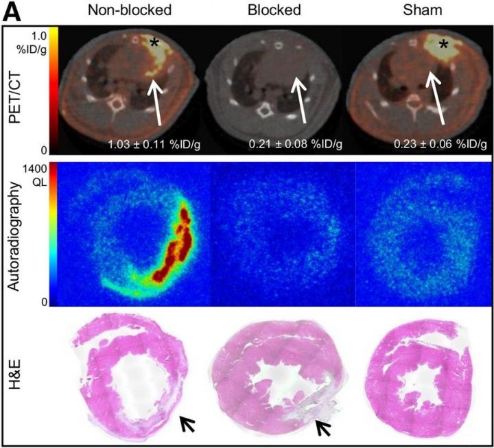
Credit: Z Varasteh and S Robu, Department of Nuclear Medicine, Klinikum rechts der Isar der TUM, Munich, Germany.
Reston, VA–Insights into how the heart recovers after myocardial infarction (heart attack) can be obtained with a new radiotracer that targets fibroblast activity prior to it causing permanent damage. According to research published in the December issue of The Journal of Nuclear Medicine, 68Ga-FAPI-04 positron emission tomography (PET) can effectively image fibroblast activation after myocardial infarction, identifying a time window during which cardiac fibrosis can be prevented and the disease course altered.
After a heart attack, fibroblasts play an important role in tissue replacement, preserving the structural integrity of the heart. However, excessive fibrosis can lead to increased left ventricle stiffness and decreased cardiac contraction. As heart failure remains a major source of late morbidity and mortality after a heart attack, study authors sought to evaluate the feasibility of imaging activated fibroblasts after myocardial infarction using a novel 68Ga-labeled fibroblast activation protein (FAP) inhibitor.
“We know that the temporospatial presence of activated fibroblasts in the injured myocardium predicts the quality of cardiac remodeling after a heart attack,” noted Zohreh Varasteh, PhD, research fellow at Klinikum rechts der Isar der TUM in Munich, Germany. “Therefore, imaging of activated fibroblasts using 68Ga-FAPI-04 PET may have significant diagnostic and prognostic value, which could aid in the clinical management of patients after myocardial infarction.”
The preclinical study included 20 rats subjected to myocardial infarction by permanent ligation of the left anterior descending coronary artery, as well as four sham-operate rats that underwent the same procedure except the ligation. In vivo imaging with 68Ga-FAPI-04 PET was performed at one, three, six, 14, 23 and 30 days after the myocardial infarction and with 18F-FDG three days after myocardial infarction. Dynamic 68Ga-FAPI-04 PET and blocking studies were performed on the myocardial infarction rats on day seven. Ex vivo imaging, autoradiography, histologic studies and immunofluorescence staining were also conducted to validate results of in vivo imaging.
Researchers found that uptake of 68Ga-FAPI-04 PET peaked six days after ligation and decreased rapidly to the background level by two weeks after infarction. Uptake accumulated mainly at the border zone of the infarcted myocardium, with high contrast and minimal uptake in normal myocardium. Minimal uptake was noted in rats that received a blocking dose of non-labeled FAPI-04. The exact location of the 68Ga-FAPI-04 uptake was confirmed by ex vivo imaging, as well as autoradiography and histologic findings.
“While preclinical development of potential anti-fibrotic approaches is far advanced, there has been little clinical validation due to the lack of sensitive and specific imaging technologies for assessing cardiac fibrosis progression or regression. In this regard, 68Ga-FAPI-04 PET has emerged as an important tool for the detection of fibrotic processes in the efforts to improve heart failure therapy,” Varasteh said.
Varasteh continued, “In the future, these advances in imaging may also be applied to other conditions associated with the activation of fibroblasts, such as hypertension, ischemic, dilated and hypertrophic cardiomyopathies, cancer therapy-related cardiotoxicity, liver cirrhosis and pulmonary fibrosis.”
###
The authors of “Molecular Imaging of Fibroblast Activity After Myocardial Infarction Using a 68Ga-Labeled Fibroblast Activation Protein Inhibitor, FAPI-04” include Zohreh Varasteh, Stephanie Robu, Miriam Braeuer, Negar Omidvari, Geoffrey Topping, Stephan G. Nekolla, Antonia Richter and Wolfgang A. Weber, Department of Nuclear Medicine, Klinikum rechts der Isar der TUM, Munich, Germany; Sarajo Mohanta and Christian Weber, Institute for Cardiovascular Prevention, Ludwig-Maximilians-Universität, Munich, Germany, and German Centre for Cardiovascular Research, Munich Heart Alliance, Munich, Germany; Yuanfang Li, Ting Sun and Andreas Habenicht, Institute for Cardiovascular Prevention, Ludwig-Maximilians-Universität, Munich, Germany; and Uwe A. Haberkorn, Department of Nuclear Medicine, University of Heidelberg, Heidelberg, Germany.
Please visit the SNMMI Media Center for more information about molecular imaging and precision medicine. To schedule an interview with the researchers, please contact Rebecca Maxey at (703) 652-6772 or [email protected]. Current and past issues of The Journal of Nuclear Medicine can be found online at http://jnm.
About The Journal of Nuclear Medicine
The Journal of Nuclear Medicine (JNM) is the world’s leading nuclear medicine, molecular imaging and theranostics journal, accessed more than 9 million times each year by practitioners around the globe, providing them with the information they need to advance this rapidly expanding field.
JNM is published by the Society of Nuclear Medicine and Molecular Imaging (SNMMI), an international scientific and medical organization dedicated to advancing nuclear medicine and molecular imaging–precision medicine that allows diagnosis and treatment to be tailored to individual patients in order to achieve the best possible outcomes. For more information, visit http://www.
Media Contact
Rebecca Maxey
[email protected]
703-652-6772
Original Source
https:/
Related Journal Article
http://dx.


