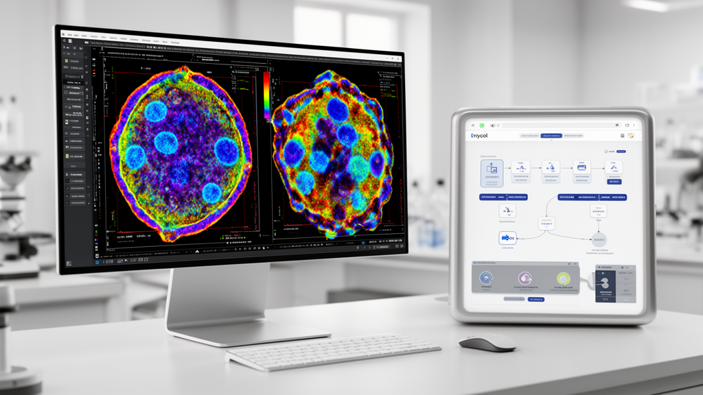
Researchers have introduced Mycol, an open-source, machine-learning-assisted image analysis platform designed to be accessible and run on standard laptops. While biological image analysis is central to modern biology, many available platforms require specialized technical expertise, code-based workflows, extensive setup, or paid access. To resolve these challenges, Mycol supports user-friendly, end-to-end machine learning workflows that incorporate automated segmentation and classification within a single interface. Users can annotate biological images, fine-tune machine learning models, inspect and correct automated predictions, and quantitatively compare morphological and class frequency descriptors. By combining automated machine-learning analysis with efficient quality control by humans, the platform makes rapid, high-quality image quantification available to non-specialists.
The platform was evaluated through three case studies spanning four distinct species to test its performance across varied biological contexts. In microbiological testing, investigators used Mycol for colony forming unit counting in Staphylococcus aureus and Escherichia coli. In agricultural pathology research, the platform evaluated spore germination in the crop pathogen Fusarium oxysporum. In marine biology investigations, the system analyzed larval development in the blue mussel Mytilus edulis. Across these diverse biological applications, researchers generated curated training sets and obtained high-quality segmentation and classification models directly within the unified software environment.
Deploying these machine learning models through Mycol produced substantial experimental efficiencies while enhancing procedural rigor. When applied to Fusarium oxysporum, using Mycol cut cell counting time by 35 percent without reducing consistency compared to established procedures. Beyond reducing analytical time, deploying these models through the platform increased the traceability of established cell counting workflows.
In addition to speeding cell counting, deploying Mycol facilitated a detailed quantitative comparison of morphological parameters that revealed new biological patterns in early Mytilus edulis larval development. Together, these results demonstrate that user-friendly machine learning workflows effectively make high-quality biological image analysis accessible on standard laptops.
Subject of Research: Development and validation of Mycol, an open-source machine learning platform for automated biological image analysis.
Article Title: Mycol: a user-friendly app for automating object identification and classification in biological images
Article References: Bradley, S. A., Webel, H., Schiesaro, G., Skumantz, M., Novillo-Sanjuan, O., Susilla, A. C., Panagou, A., Lucena-Marín, R., Whitford, C. M., Jensen, E. D., Di Pietro, A., & Acevedo-Rocha, C. G. (2026). Mycol: a user-friendly app for automating object identification and classification in biological images. Applied Microbiology and Biotechnology. https://doi.org/10.1007/s00253-026-14097-1
Image Credits: AI Generated
DOI: 10.1007/s00253-026-14097-1
Keywords: Biological image analysis, Machine learning, Mycol, Open-source software, Automated segmentation, Image classification, Staphylococcus aureus, Escherichia coli, Fusarium oxysporum, Mytilus edulis, Larval development, Colony forming units, Applied microbiology, Biotechnology, User-friendly workflows, Quantitative microscopy
News Source: Gregory Coleman. (November 19, 2026). New Open-Source App Mycol Accelerates Biological Image Analysis and Quantification. Scienmag.