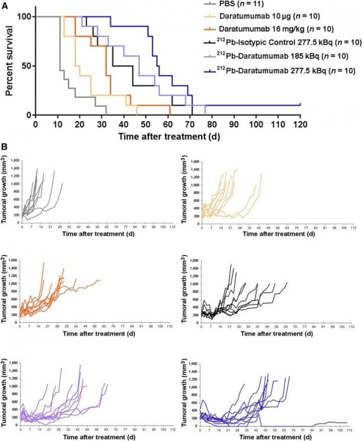
Credit: Images created by S. Durand-Panteix and I. Quelven-Bertin, et al, University of Limoges, CNRS, INSERM
Reston, VA–A new alpha-radioimmunotherapy, 212Pb-anti-CD38, has proven effective in preventing tumor growth and increasing survival in multiple myeloma tumor-bearing mice, according to new research published in the July issue of the Journal of Nuclear Medicine. Given the long half-life, central production and worldwide distribution of 212Pb-anti-CD38, researchers have determined that the α-radioimmunotherapy is not only effective but also clinically feasible as a multiple myeloma treatment.
Multiple myeloma is a plasma cell cancer that occurs in bone marrow. It is the second most common type of blood cancer, with more than 32,000 new cases projected in 2020, according to the American Cancer Society. Despite new treatments and protocols, the prognosis of multiple myeloma patients remains poor, as remission is often followed by relapse. Innovative therapies with a distinct mechanism of action are therefore needed.
“Targeted radioimmunotherapy using antibodies against proteins expressed at the cell membrane on tumoral cells has already shown efficacy with beta-emitters, but with severe side effects,” said Isabelle Quelven-Bertin, PharmD, radiopharmacist at Limoges University Hospital in Limoges, France. “Using alpha particles is very attractive because their much shorter path-length reduces unwanted radiation exposure on normal tissues, thereby reducing side effects. As such, we investigated the potential of the α-radioimmunotherapy 212Pb-anti-CD38 in the treatment of multiple myeloma.”
In the study, researchers developed human myeloma cell lines, which were analyzed for cell proliferation after incubation with various concentrations of 212Pb monoclonal antibodies. Mice received subcutaneous grafts of human myeloma cell lines and were injected with 212Pb-anti-CD38, 212Pb-anti-mCD38 (mice-specific) or 212Pb-isotypic control. Biodistribution, toxicity and dose-range-finding studies were performed, as well as radioimmunotherapy experiments that measured tumor volume and overall survival.
The human myeloma cell line saw a significant inhibition of cell proliferation three days after incubation with 212Pb-anti-CD38. Likewise, the mouse model showed marked inhibition of cell proliferation for mice treated with 212Pb-anti-CD38, with a median survival of 55 days; mice that received the isotypic control had a median survival of 11 days. Biodistribution studies showed a specific tumoral accumulation of 212Pb-anti-CD38, and toxicity experiments with 212Pb-anti-mCD38 established a toxic activity of 277.5 kBq.
While 212Pb monoclonal antibodies effectively inhibited tumor growth and increased survival, whole-body SPECT/CT imaging was not possible due to the low 212Pb activity injected and the detection sensitivity. Instead, researchers performed SPECT/CT imaging with 203Pb, a chemically identical radiometal.
“This radioelement allowed us to consider a theranostic approach for 212Pb α-radioimmunotherapy, preventing the need for a radionuclide with different physical-chemical properties that would likely result in different pharmacokinetics. Combining therapy and imaging could provide an effective approach to optimizing therapeutic doses using specific dosimetry calculation and to monitoring the patient’s response. Moving forward, this could be considered as an innovative theranostic approach,” noted Michel Cogné, MD, PhD, professor of immunology at Limoges Medical School in Limoges, France.
###
This study was made available online in December 2019 ahead of final publication in print in July 2020.
The authors of “212Pb α-Radioimmunotherapy Targeting CD38 in Multiple Myeloma: A Preclinical Study” include Isabelle Quelven and Jacques Monteil, Nuclear Medicine Department, Limoges University Hospital, Limoges, France, and CNRS-UMR7276, INSERM U1262, Contrôle de la Réponse Immune B et Lymphoproliférations, Limoges University, Limoges, France; Audrey Bayout, Nuclear Medicine Department, Limoges University Hospital, Limoges, France; Magali Sage, Jérémy Mounier, Julie Garrier, Michel Cogné and Stéphanie Durand-Panteix, CNRS-UMR7276, INSERM U1262, Contrôle de la Réponse Immune B et Lymphoproliférations, Limoges University, Limoges, France; and Amal Saidi, Orano Med SAS, Paris, France.
Please visit the SNMMI Media Center for more information about molecular imaging and precision imaging. To schedule an interview with the researchers, please contact Rebecca Maxey at (703) 652-6772 or [email protected].
About the Society of Nuclear Medicine and Molecular Imaging
The Journal of Nuclear Medicine (JNM) is the world’s leading nuclear medicine, molecular imaging and theranostics journal, accessed close to 10 million times each year by practitioners around the globe, providing them with the information they need to advance this rapidly expanding field. Current and past issues of The Journal of Nuclear Medicine can be found online at http://jnm.
JNM is published by the Society of Nuclear Medicine and Molecular Imaging (SNMMI), an international scientific and medical organization dedicated to advancing nuclear medicine and molecular imaging–precision medicine that allows diagnosis and treatment to be tailored to individual patients in order to achieve the best possible outcomes. For more information, visit http://www.
Media Contact
Rebecca Maxey
[email protected]
Original Source
http://www.
Related Journal Article
http://dx.


