


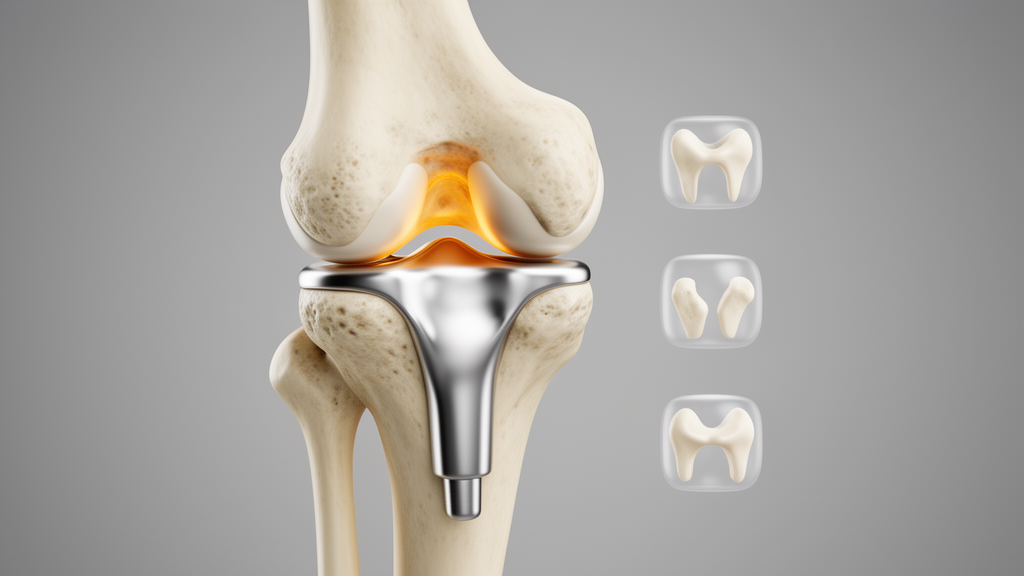
Orthopedic researchers have introduced a four-type classification system for anterior femoral notching, a common and often debated phenomenon in total knee arthroplasty, and preliminary testing suggests surgeons can apply it with substantial agreement. The Femoral Notch Classification, published in the journal Arthroplasty, sorts the geometric relationship between the anterior femoral resection plane, the native cortical line, and the anterior flange of the femoral component into four descriptive categories, from complete cortical preservation to a critical notch exceeding three millimeters. In a reliability study of 80 postoperative lateral radiographs, three independent observers reached an inter-observer Fleiss kappa of 0.80 and a quadratic weighted kappa of 0.87, while intra-observer agreement measured by Cohen’s kappa averaged 0.91, in the almost perfect range.
Anterior femoral notching occurs when the saw cut across the front of the distal femur removes bone below the level of the native anterior cortex. The practice has historically been viewed primarily as a risk factor for postoperative supracondylar periprosthetic fracture, and large clinical series have reported fracture prevalence and outcomes after notching. Biomechanical studies cited by the authors found that notches deeper than 3 millimeters can reduce distal femoral strength by roughly 18 to 31 percent, particularly when cortical integrity is compromised or bone quality is reduced. More recent clinical data, however, indicate that not every notch correlates with a higher fracture incidence; the risk appears to be modulated by notch depth, cortical involvement, and patient factors rather than by the mere presence of a notch.
The authors, Alexander Antoniadis, Jaad Mahlouly, Thibaut Royon and Julien Wegrzyn, argue that notching also matters well beyond fracture risk. Femoral notching influences implant fit, patellofemoral balance, joint stability, and overall limb biomechanics. Computational finite element analyses and gait simulations show that notch depth alters stress distribution in the distal femur during the gait cycle, which underscores the need to coordinate anterior and posterior resections so that neither compartment is over- or under-resected. Techniques that restore distal femoral anatomy with minimal compromise, such as anatomic or hybrid referencing, aim to balance the anterior and posterior gaps, reduce unintended cortical violation, and optimize patellofemoral mechanics.
Despite this clinical importance, the team notes that no widely adopted, radiograph-based classification has been dedicated to anterior femoral notching in knee replacement. The Tayside scheme, which grades postoperative notching into three levels according to the depth and extent of cortical and cancellous involvement, remains the most recognized system, but it offers limited intraoperative guidance, does not capture subtler radiographic patterns, and has not achieved widespread adoption. Many reports still describe notching qualitatively as present or absent, or mild, moderate or severe, without a standardized framework. The new classification was therefore designed as a descriptive, radiograph-based common language intended to work both intraoperatively, independent of implant design or alignment philosophy, and on postoperative lateral radiographs.
The four categories are anchored to standard anteroposterior and lateral radiographs obtained according to the Modern Knee Society radiographic methodology. Type 0, called No Notch, describes complete preservation of the anterior cortex with no visible step-off or indentation; the anterior resection may match the femoral component thickness precisely, and if it does not, anterior overstuffing or posterior over-resection must be controlled. Type 1, On the Line, describes an anterior resection flush with the femoral cortex, producing a subtle notch appearance that remains biomechanically stable without compromising the cortex. Type 2, the Controlled Notch, involves a deeper resection, ideally less than 3 millimeters, beyond the cortical line, with a smooth contour and preserved cortical continuity, and is considered acceptable when the radiographic pattern is consistent with accommodation of the native trochlear geometry. Type 3, the Critical Notch, is an excessive resection of more than 3 millimeters with significant cortical interruption and no radiographically identifiable anatomical correlate, a configuration that may compromise distal femoral strength and is associated with increased periprosthetic fracture risk when not justified by trochlear geometry.
The distinction between a controlled and a critical notch rests on notch depth measured from the anterior cortical line on a true lateral radiograph, preservation of cortical continuity, and comparison with preoperative radiographs to assess the underlying trochlear geometry. Notch depth is defined as the maximum perpendicular distance between the anterior cortical line and the anterior femoral cut, and a true lateral projection with overlapping posterior condyles is required for reliable measurement. The 3-millimeter threshold serves as a practical reference point drawn from previous biomechanical studies, although the authors emphasize that classification is based primarily on qualitative radiographic patterns rather than absolute measurements, with cortical continuity and preoperative comparison also incorporated.
The concept emerged from clinical observation that postoperative lateral radiographs show reproducible patterns of anterior femoral morphology after knee replacement, ranging from extensive anterior cortical loss to a smooth cortical profile compatible with the patient’s native anatomy. Common indications for a deliberately controlled notch include variations in distal femoral anatomy, such as pronounced anterior curvature, a shallow or dysplastic trochlea, or a mismatch between native trochlear geometry and implant design, situations that would otherwise lead to anterior overstuffing or compromised patellofemoral tracking if a notch were strictly avoided. The authors stress that the biomechanical rationale is drawn from previously published literature and was not newly evaluated in this study, which focused on classification development and preliminary reliability testing.
For the reliability assessment, three observers of different experience levels, one senior orthopedic surgeon and two fellows, classified 80 anonymized true lateral radiographs from primary total knee arthroplasties performed at a single institution, all meeting Modern Knee Society standards. Observers were blinded to clinical information and to each other’s ratings, and after four weeks the same set was presented in randomized order to measure intra-observer agreement. Notably, the observers had access to the corresponding preoperative radiographs to evaluate native trochlear geometry when classifying the postoperative images. Most radiographs fell into Type 0 and Type 1, with only 10 Type 2 cases and a single Type 3 case, a distribution the authors themselves flag as a key limitation.
That limitation matters for interpretation. Because agreement was largely driven by the frequent Types 0 and 1, reliability estimates for Types 2 and 3 should be regarded as preliminary pending larger external validation. The study was conducted at a single institution, distinguishing Type 2 from Type 3 can be difficult when notch depth approaches 3 millimeters or cortical continuity is borderline, and radiographic assessment of anatomic context may be inherently subjective. Although the classification is designed to be applicable intraoperatively, the reliability analysis was performed exclusively on postoperative images, so its intraoperative utility remains a proposed application requiring further validation. Implant- and technique-agnostic in principle, the system could still be influenced by subtle differences in trochlear flange design across manufacturers.
The authors position the classification as a practical step toward standardized reporting in research and registry data, potentially supporting more consistent outcome studies than current qualitative or depth-only descriptions. They suggest that future investigations should assess external validity and correlations with clinical outcomes, including supracondylar fracture rates, anterior knee pain, patellofemoral complications, and implant survival. By distinguishing anatomically appropriate configurations from those potentially compromising cortical integrity, the framework may reduce unnecessary concern over controlled resections while maintaining vigilance against excessive cortical violation. For now, the system is promising in concept but prospective validation of its reliability and clinical impact remains outstanding, and the authors recommend consulting preoperative radiographs whenever distinguishing a controlled from a critical notch proves difficult.
Subject of Research: A new four-type radiographic classification system for anterior femoral notching in total knee arthroplasty and its preliminary reliability testing.
Article Title: A novel classification system for anterior femoral notching in total knee arthroplasty
Article References: Antoniadis, A., Mahlouly, J., Royon, T., & Wegrzyn, J. (2026). A novel classification system for anterior femoral notching in total knee arthroplasty. Arthroplasty, 8(1), Article 66. https://doi.org/10.1186/s42836-026-00424-4
Image Credits: AI Generated
DOI: 10.1186/s42836-026-00424-4
Keywords: total knee arthroplasty, femoral notching, classification system, periprosthetic fracture, radiographic assessment, inter-observer reliability, trochlear geometry, anterior cortex, kappa statistic, orthopedic surgery, novel, classification
News Source: Ophelia Keating. (October 30, 2026). New Four-Type System Classifies Femoral Notching in Knee Replacement. Scienmag.



