• HOME
  • NEWS
  • EXPLORE
    • CAREER
      • Companies
      • Jobs
    • EVENTS
    • iGEM
      • News
      • Team
    • PHOTOS
    • VIDEO
    • WIKI
  • BLOG
  • COMMUNITY
    • FACEBOOK
    • INSTAGRAM
    • TWITTER
Sunday, November 9, 2025
BIOENGINEER.ORG
No Result
View All Result
  • Login
  • HOME
  • NEWS
  • EXPLORE
    • CAREER
      • Companies
      • Jobs
        • Lecturer
        • PhD Studentship
        • Postdoc
        • Research Assistant
    • EVENTS
    • iGEM
      • News
      • Team
    • PHOTOS
    • VIDEO
    • WIKI
  • BLOG
  • COMMUNITY
    • FACEBOOK
    • INSTAGRAM
    • TWITTER
No Result
View All Result
Bioengineer.org
No Result
View All Result
Home NEWS Science News Biology

New fluorescence method reveals signatures of individual microbes

Bioengineer by Bioengineer
October 4, 2019
in Biology
Reading Time: 2 mins read
0
IMAGE
Share on FacebookShare on TwitterShare on LinkedinShare on RedditShare on Telegram

Researchers from University of Tsukuba develop a new method that reveals the unique signatures of individual cells in mixtures of bacteria, fungi and yeast under a microscope

IMAGE

Credit: University of Tsukuba

Tsukuba, Japan – When viewed with specialized microscopes, microbial cells show an individual fluorescence pattern, or signature, that depends on the mixture of biomolecules contained within the cells. That complex mixture, with its telltale signature, in turn depends on the type of microbe and its physiological state, such as whether it is growing or what it is consuming as a food source.

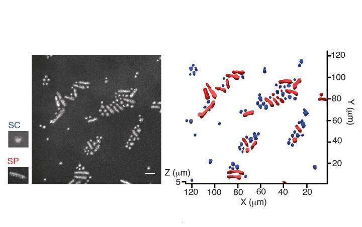
University of Tsukuba researchers have developed a new method, named CRIF (Confocal Reflection microscopy-assisted single-cell Innate Fluorescence), to detect the fluorescence signatures of individual microbial cells. The method is non-destructive, meaning the cells remain alive, and allows cells to be studied in realistic three-dimensional environments. Importantly, the new method can be used to view individual cells within mixtures of different types of microbe, unlike many standard techniques that work best with “pure” populations where the cells are all alike. The team recently published their findings in Applied and Environmental Microbiology.

“We used a confocal microscope, which can generate images from three-dimensional materials–rather like comparing a sequence of slices to build up an image of a whole intact structure. This enabled us to find the locations of the microbial cells within the sample,” says lead and co-corresponding author Yutaka Yawata. “Combining this with spectroscopy to measure a set of fluorescence signals under the microscope, we were able to see the different types of microbes within the sample, and read their signatures to identify the individual cells.”

The team went on to train computer models to read the fluorescence signatures and distinguish different types of cells. The models learned to recognize cells automatically, even when looking at different cells with very similar shapes and sizes, and analyze the images for cell signatures to identify the cells according to their type and physiological state.

“Our technique to recognize and track the innate fluorescence signatures of each of the individual cells within three-dimensional samples opens up exciting new opportunities to explore mixed microbial populations, as found in natural environments,” says co-corresponding author Nobuhiko Nomura. “It will help researchers understand how microbes grow and interact with each other in the real world.”

###

Media Contact
Naoko Yamashina
[email protected]

Related Journal Article

http://dx.doi.org/10.1128/AEM.00608-19

Tags: BacteriologyBiologyMicrobiologyMycology
Share12Tweet8Share2ShareShareShare2

Related Posts

First Hybrid Eriocheir Discovery in Mediterranean Sea

First Hybrid Eriocheir Discovery in Mediterranean Sea

November 9, 2025
Unveiling Extended-Core Gene Variation in E. coli Pan-genome

Unveiling Extended-Core Gene Variation in E. coli Pan-genome

November 8, 2025

Exploring Spanish Roma’s Genetic Diversity and Structure

November 8, 2025

Single-Cell Insights into Bat Viral Infections Uncovered

November 8, 2025
Please login to join discussion

POPULAR NEWS

  • blank

    Stinkbug Leg Organ Hosts Symbiotic Fungi That Protect Eggs from Parasitic Wasps

    314 shares
    Share 126 Tweet 79
  • ESMO 2025: mRNA COVID Vaccines Enhance Efficacy of Cancer Immunotherapy

    207 shares
    Share 83 Tweet 52
  • New Study Suggests ALS and MS May Stem from Common Environmental Factor

    139 shares
    Share 56 Tweet 35
  • Sperm MicroRNAs: Crucial Mediators of Paternal Exercise Capacity Transmission

    1302 shares
    Share 520 Tweet 325

About

We bring you the latest biotechnology news from best research centers and universities around the world. Check our website.

Follow us

Recent News

Ellagic Acid Shields Tooth Adhesives Post-Bleaching

First Hybrid Eriocheir Discovery in Mediterranean Sea

Assessing Social Anxiety in Autism: A Multi-Method Approach

Subscribe to Blog via Email

Enter your email address to subscribe to this blog and receive notifications of new posts by email.

Join 69 other subscribers
  • Contact Us

Bioengineer.org © Copyright 2023 All Rights Reserved.

Welcome Back!

Login to your account below

Forgotten Password?

Retrieve your password

Please enter your username or email address to reset your password.

Log In
No Result
View All Result
  • Homepages
    • Home Page 1
    • Home Page 2
  • News
  • National
  • Business
  • Health
  • Lifestyle
  • Science

Bioengineer.org © Copyright 2023 All Rights Reserved.