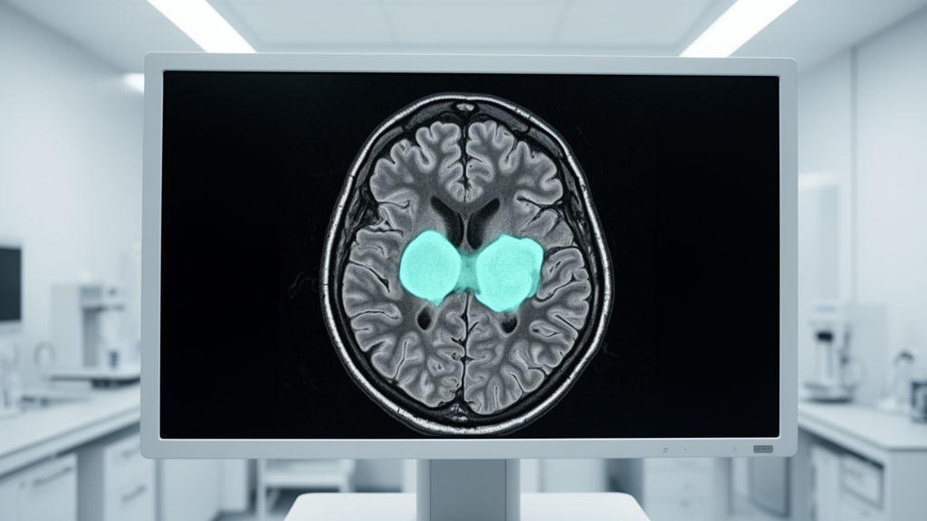
Researchers have developed an efficient, FLAIR-only deep learning framework designed to improve the accuracy of brain tumor segmentation and provide explainable survival analysis. This study, published in the International Journal of Computational Intelligence Systems, addresses a major limitation in existing medical imaging models, which often rely on complex, computationally demanding, multi-modal 3D MRI acquisitions that can hinder clinical implementation.
The proposed architecture utilizes a 2.5D contextual segmentation design that processes five-slice 2.5D FLAIR neighborhoods. By employing an Adaptive Inter-Slice Context Fusion (AICF) module, the system learns content-dependent slice relevance before projecting the data through an ImageNet-pretrained ResNet50 encoder. This approach improves volumetric context modeling without the need for traditional, resource-intensive 3D convolutions.
For the reconstruction of multiscale features, the researchers implemented an attention-modulated UNet++ Lite decoder. To handle the challenges of severe class imbalance in tumor data, the team integrated a hybrid loss function that combines categorical cross-entropy with focal Tversky loss. Additional techniques, including test-time augmentation and conditional largest-connected-component filtering, were utilized to ensure prediction consistency and robustness.
The performance metrics for the AICF-enabled model demonstrate high accuracy in tumor delineation. The system achieved a whole-tumor Dice score of 0.899 and an intersection-over-union (IoU) of 0.817. Sensitivity was measured at 0.910, with a Surface Dice at 3 mm of 0.968 and a physical-space Hausdorff distance (HD95) of 0.96 mm. Furthermore, the model showed strong voxel-wise performance, with PR-AUC at 0.895 and ROC-AUC reaching 0.9988.
Beyond raw segmentation metrics, the researchers conducted temperature-scaled calibration and uncertainty analyses. These evaluations showed that the model maintains improved agreement between its confidence levels and actual outcomes.
For prognostic evaluation, the researchers constructed a primary interpretable baseline using age and verified resection status through Kaplan–Meier estimation and penalized Cox proportional-hazards modeling. A secondary exploratory analysis was conducted to determine if specific tumor burden and morphology descriptors, derived from the segmentation process, could provide incremental prognostic information. Using apriori association-rule mining, the team identified clinically intuitive age-based risk strata with distinct survival trajectories.
Despite these results, the study has significant limitations. The findings are based on an internal cohort from the BraTS 2019 dataset and lack independent external validation. The researchers emphasize that these results should be interpreted as benchmark-level evidence rather than a demonstration of clinical deployment readiness.
Subject of Research: Computational Neuro-oncology and Medical Imaging AI
Article Title: An Efficient FLAIR-Only Deep Learning Framework for Brain Tumour Segmentation and Explainable Survival Analysis
Article References: Wankhede, D. S., Hamid, N. A. W. A., Sunder, R., Dafik, D., Phadke, M., & Deshmukh, P. V. (2026). An Efficient FLAIR-Only Deep Learning Framework for Brain Tumour Segmentation and Explainable Survival Analysis. International Journal of Computational Intelligence Systems. https://doi.org/10.1007/s44196-026-01694-1
Image Credits: AI Generated
DOI: 10.1007/s44196-026-01694-1
Keywords: Brain tumor segmentation, Deep learning, FLAIR MRI, Image processing, Survival analysis, ResNet50, UNet++, Medical AI, Radiology, Neuro-oncology, Computational intelligence, Clinical research, Prognostic assessment, Predictive modeling, Adaptive Inter-Slice Context Fusion
News Source: Nathaniel Bowman. (November 16, 2026). New FLAIR-Only AI Model Achieves Precise Brain Tumor Segmentation Accuracy. Scienmag.