


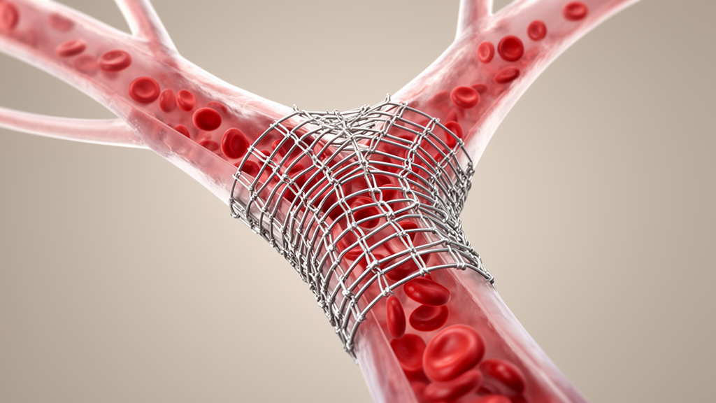
Surgeons have long faced an awkward problem at the top of the chest: when a tumour or scar tissue squeezes the superior vena cava and the two brachiocephalic veins that feed it, the standard endovascular fixes each carry a price. A team of Italian interventional radiologists and vascular specialists now describes a way to rebuild that entire venous junction with covered stents while keeping both arms’ drainage pathways open, a configuration they call Covered Endovascular Reconstruction of the Anonymous Confluence, or CERAC. In a technical note published in CVIR Endovascular, the group from Policlinico San Matteo in Pavia reports that the technique achieved technical success in all three patients in whom it was used, with no immediate complications and patent stents throughout follow-up periods ranging from 29 to 301 days.
The superior vena cava is the large vein that collects blood from the head, neck and both arms and delivers it to the heart. When something blocks it, the result is superior vena cava syndrome: swelling of the face and arms, distended neck veins, shortness of breath and, in severe cases, airway compromise. Malignant causes remain common, but the authors note that benign cases have been climbing in recent years, driven largely by thrombosis around long-term central venous access devices such as catheters and ports. Those benign patients tend to be younger and to face decades of life ahead, which makes durable venous patency and preservation of future vascular access for dialysis, chemotherapy or monitoring particularly important.
Endovascular therapy, in which stents are delivered through a vein in the groin rather than by open surgery, is the accepted first-line treatment for the syndrome. Yet the optimal strategy for lesions that involve the brachiocephalic confluence, the Y-shaped junction where the right and left brachiocephalic veins merge into the superior vena cava, remains contested. Most published studies favour stenting only one side, because unilateral reconstruction relieves symptoms acceptably and carries lower complication rates. Bilateral stenting is generally reserved for selected cases in which the confluence itself and the cava are both involved, or when the cava’s diameter exceeds 15 millimetres.
The existing bilateral options each have documented drawbacks. Kissing or double-barrel configurations place two parallel stents side by side inside the cava, a geometry that leaves gaps in contact with the vessel wall, promotes leakage around the stents and creates turbulent flow at the midline seam where blood stagnates and clots may form. Y-stenting requires a branch limb to be threaded through the struts of the cava stent, which is structurally incompatible with a covered cava stent. The train-track technique does achieve anatomical reconstruction of the confluence, but with bare stents, whose open mesh allows tumour to grow between the struts, a failure mode reported in up to roughly 40 percent of malignant cava stenting cases. The train-track authors themselves acknowledge technical constraints, including the need for buddy-wire dilatation and the impossibility of using stents or balloons with long tapered tips.
CERAC was conceived to sidestep those limitations. The concept borrows from the Covered Endovascular Reconstruction of the Aortic Bifurcation, a technique used in the aorta, and adapts it to the venous junction. In the illustrative case, a 26-year-old man with a mixed germ cell mediastinal tumour presented with facial and chest swelling and a swollen right arm; computed tomography showed a large anterior mediastinal mass encasing the brachiocephalic veins and compressing the cava. Because preserving both venous pathways mattered for his future treatment, the team chose CERAC. The authors report that the procedure begins with antibiotic prophylaxis, 5000 international units of unfractionated heparin, and two weeks of prophylactic low-molecular-weight heparin afterwards; no routine antiplatelet therapy is given, reflecting the unproven role of antiplatelet drugs in venous circulation and the large stent diameters used under brisk venous flow.
Under local anaesthesia and conscious sedation with midazolam, a single ultrasound-guided femoral vein access is established with a 6-French sheath. A hydrophilic guidewire and catheter navigate through the stenotic or occluded segments, and venography maps the anatomy. The wire is then exchanged for a stiff guidewire, and an 18-French, 65-centimetre long sheath is introduced to accommodate the covered stent systems. The contralateral brachiocephalic vein is accessed the same way until stiff guidewires sit in both stenotic brachiocephalic veins. A 16-millimetre covered stent is then deployed in the superior vena cava over one of the wires, extending from just below the confluence to the proximal cava so that the obstructed segment is completely covered.
Before removing the stiff guidewire jailed between the caval stent and the vessel wall, the ipsilateral brachiocephalic vein is selectively reaccessed through the caval stent with a hydrophilic guidewire and catheter; only then is the jailed wire carefully withdrawn and exchanged for a stiff wire. The caval stent is post-dilated with a balloon sized to match or slightly undersize the nominal stent diameter to optimise sealing and expansion. Two 12-millimetre covered stents are then deployed in the brachiocephalic veins. The first is advanced, released and post-dilated; while its angioplasty balloon remains inflated, the second stent is advanced into the contralateral vein to avoid impingement or displacement during positioning. After balloon deflation the second stent is deployed, and both are post-dilated simultaneously with the kissing balloon technique. Each brachiocephalic stent is positioned to extend roughly one centimetre into the cava stent, minimising overlap while maintaining complete coverage, and distal elongation can be performed if required.
The result is a so-called double-D-shaped lumen at the level of the confluence, in which the caudal ends of the brachiocephalic stents flare within the larger cava stent. Final venography confirms patency of both brachiocephalic veins and the cava, and a post-procedural cone-beam CT can be obtained to assess stent apposition and spatial configuration at different anatomical levels. Haemostasis is achieved with a purse-string suture. The authors argue that this geometry preserves a larger lumen and minimises excluded space compared with parallel-barrel designs, and that the interlocking configuration may also guard against late stent migration, a concern they note has not been reported in the principal published series of covered stenting for malignant cava obstruction.
Between September 2024 and December 2025, ten patients with superior vena cava syndrome underwent endovascular treatment at the institution. CERAC was selectively applied in three. In two young patients the goal was to preserve bilateral upper-extremity venous access in the setting of potentially treatable malignant mediastinal disease. In one patient the procedure was performed emergently to achieve rapid central venous decompression because severe upper-airway oedema prevented safe removal of a tracheal cannula. The remaining seven patients received unilateral reconstruction. Mean fluoroscopy time across the three CERAC cases was 30.6 minutes. Final angiography showed restoration of central venous patency in all patients, with no immediate procedural complications, and all patients experienced rapid clinical improvement with resolution of venous congestion symptoms. Primary stent patency was maintained throughout available follow-up, which ranged from 29 to 301 days with a median of 149 days.
The authors also address a potential objection: covering the azygos vein’s opening into the cava. In principle this is to be avoided, but they argue that in patients with extensive central venous obstruction involving both the confluence and the cava, the azygos system is typically already non-functional as a collateral or is working in retrograde direction towards the inferior vena cava, so once antegrade cava flow is restored its collateral role becomes physiologically redundant. They report that no clinically relevant adverse event attributable to azygos jailing has appeared in published series of unilateral or bilateral covered cava stenting, and none of their own patients developed signs or symptoms attributable to azygos occlusion during follow-up. The team is candid about the limits of the report: the sample is small, follow-up is heterogeneous, no follow-up CT documenting significant tumour regression was available, and the haemodynamic advantage hypothesised for the double-D configuration rests on indirect arterial evidence rather than on flow modelling or imaging in the venous setting. The anti-migration rationale has likewise not been specifically validated in the context of tumour regression after systemic therapy. As a Level V technical note, the work establishes feasibility and reproducibility in selected cases rather than comparative superiority, and the authors call for further studies to assess long-term patency and to refine patient selection.
Subject of Research: A covered-stent endovascular technique for reconstructing the brachiocephalic vein confluence in superior vena cava syndrome
Article Title: Covered Endovascular Reconstruction of the Anonymous Confluence (CERAC): a technical note describing a confluence-preserving endovascular technique
Article References: Corti, R., Ambrosi, L., Cionfoli, N., D’Agostino, M. A., Quaretti, P., & Bozzani, A. (2026). Covered Endovascular Reconstruction of the Anonymous Confluence (CERAC): a technical note describing a confluence-preserving endovascular technique. CVIR Endovascular, 9(1), Article 119. https://doi.org/10.1186/s42155-026-00765-w
Image Credits: AI Generated
DOI: 10.1186/s42155-026-00765-w
Keywords: superior vena cava syndrome, CERAC, covered stents, brachiocephalic confluence, endovascular reconstruction, central venous access, venous stenting, technical note, case series, interventional radiology, stent patency, mediastinal tumour
News Source: Ophelia Keating. (October 30, 2026). New CERAC Stent Technique Rebuilds the Vein Junction to Restore Blood Flow. Scienmag.



