Credit: Mika Reinisalo
Researchers at the University of Eastern Finland have developed two new cell models that can open up new avenues for ocular drug discovery. The new cell models are continuously growing retinal pigment epithelial cells, which have many benefits over the models currently used by researchers and pharmaceutical companies. The models were developed by Professor Arto Urtti’s Ocular Drug Delivery group at the University of Eastern Finland.
The retinal pigment epithelium is located in the back of the eye, forming the outer blood-retinal barrier. Pigment epithelial cells play a key role in, e.g., age-related macular degeneration, which makes them an interesting target for drug therapy. The retinal pigment epithelium also regulates the access of drugs from the blood stream into the eye and vice versa, further highlighting the importance of this cell type for drug discovery.
Up until now, cultivated retinal pigment epithelial cell lines have lacked both pigmentation and the blood-retinal barrier, which has complicated their use.
Cells can be fed with pigment that binds drugs
The retinal pigment epithelium (RPE) is characterised by strong pigmentation. Many drugs bind to cellular pigment and, consequently, can accumulate in RPE cells. The new cell model makes it possible to study this accumulation in greater detail than before.
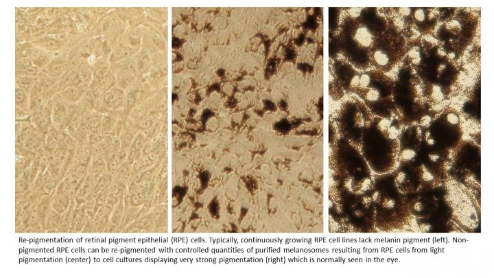
In the new model, non-pigmented RPE cells can be re-pigmented by feeding them with melanin pigment.
“Re-pigmentation is possible, thanks to the naturally active phagocytosis, or cellular eating, of RPE cells. Instead of just using pigment, we used melanosomes, i.e. live pigmented cell organelles, to re-pigment the cells. Our research group has already previously developed a method to isolate live melanosomes. In this study, we showed that the host cell accepts the transferred melanosomes and doesn’t destroy them,” Postdoctoral Researcher Mika Reinisalo from the University of Eastern Finland says.
Published in Scientific Reports, the study also described the uptake of different drugs in melanosomes. According to the researchers, most drugs that bind to melanin at high levels were taken up by the melanosomes, whereas the same was true only for a small number of drugs that bind to melanin at low levels.
“By using non-pigmented cells as controls, we were able to study how much of a drug given to the cells eventually ends up in the melanosomes. Next, we need to find out for how long the melanosomes can hold on to a drug, and whether this causes any harm. The accumulation of drugs in the melanosomes can, on the other hand, make it possible to target drugs at specific tissues,” Reinisalo notes.
New cell model forms a more realistic blood-retinal barrier
Retinal pigment epithelial cells also form the blood-retinal barrier that protects the eye from circulating xenobiotics. For drug therapy, the blood-retinal barrier is crucial, as it regulates both the entry and exit of drugs to and from the eye. In earlier cell models, the epithelial layer isn’t thick enough, allowing drugs to pass through it too quickly. This paints an erroneous picture of how drugs get transported into the patient’s eye.
Earlier this year, the same research group published a study in Pharmaceuticals, presenting a new RPE cell population that had spontaneously arisen from the RPE cell line. This population forms a tight epithelial layer, resembling the real-life epithelial layer of the eye.
“We named this cell population after its discoverer, Senior Laboratory Technician Lea Pirskanen, who is also one of the authors of the study. These cells are now known as LEPI cells. We decided to study them in more detail and discovered that in comparison to earlier cell models, LEPI cells are better differentiated and they form a tighter and more realistic barrier through which drugs have to pass,” Postdoctoral Researcher Laura Hellinen from the University of Eastern Finland says.
Both cell models are useful for ocular drug discovery and they also reduce the need for animal testing.
###
The studies were funded by the Academy of Finland and the Finnish Cultural Foundation.
For further information, please contact:
Postdoctoral Researcher Mika Reinisalo, University of Eastern Finland, [email protected]
Ocular Drug Delivery Research Group at the University of Eastern Finland:
https:/
Research articles:
Hellinen Laura, Hagström Marja, Knuutila Heidi, Ruponen Marika, Urtti Arto and Reinisalo Mika. Characterization of artificially re-pigmented ARPE-19 retinal pigment epithelial cell model. Scientific Reports, vol. 9, Article number: 13761 (2019).
https:/
Hellinen Laura, Pirskanen Lea, Tengvall-Unadike Unni, Urtti Arto and Reinisalo Mika. Retinal pigment epithelial cell line with fast differentiation and improved barrier properties. Pharmaceutics 2019, 11(8), 412.
https:/
Media Contact
Mika Reinisalo
[email protected]
Related Journal Article
http://dx.


