Credit: T. Usui / TUAT
A research team led by scientists from Tokyo University of Agriculture and Technology (TUAT), Japan, has successfully established 3D cultured tissue that mimics liver fibrosis, a key characteristic of non-alcoholic steatohepatitis (NASH). For making the 3D culture, cells were collected from liver tissues of NASH model mice. Their findings open up an alternative avenue for developing drugs for NASH patients, identifying new markers for early diagnosis, and better understanding the disease progression.
Their findings were published in Biomaterials on Jan 27th, 2020.
In Japan, about 10 million of people (8% of the population) are thought to carry NASH or to be at high risk for NASH. In the USA, a ballpark estimate indicates about 3% to 12% of adults have NASH. The NASH patients develop fatty liver regardless of alcohol intake and progress to liver cirrhosis or ultimately liver cancer. NASH symptoms include liver tissue inflammation, fat deposition, and fibrosis. Unfortunately, there are no medications available for treating NASH, thus this becomes a big social problem.
In general, to test which drugs could cure NASH, researchers used the method that experimental animals were fed by a NASH-inducing diet, followed by drugs were continuously given to these animals. It is, however, obvious that using experimental animals to test drugs is not practical. “To mimic NASH in a dish, we started three-dimensionally growing cells that were collected from liver tissues of NASH model mice”, said Dr. Tatsuya Usui, corresponding author of the paper, Senior Assistant Professor, Laboratory of Veterinary Pharmacology, Department of Veterinary Medicine, Faculty of Agriculture, TUAT. “These cells successfully grew in a dish and formed mini-organs called organoids.”
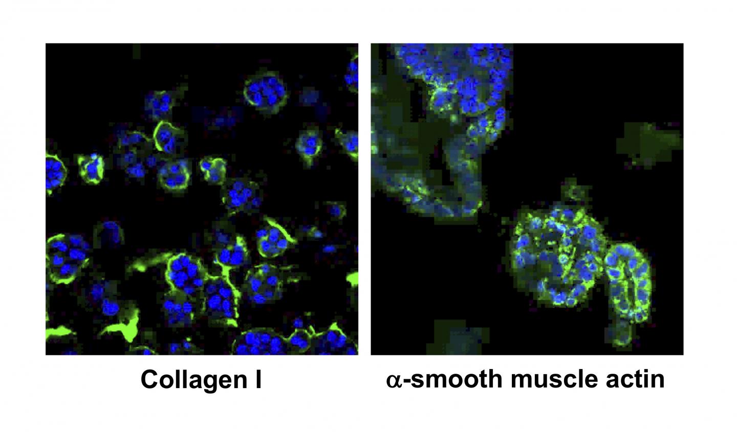
For producing NASH organoids in a dish, we used cells from liver in the NASH mice with 3 different disease stages, such as the early stage (fatty liver), the middle stage (fatty liver), and the late stage (advanced fibrosis). The organoids were examined by standard histology methods, such as HE staining, oil red staining, and Masson’s trichrome staining to visualize cell shapes, oil production in cells, and connective issues, respectively. In addition, immuno-staining, quantitative PCR, RNA-sequencing were performed to see localization and amount of bio-markers.
“After careful scientific analyzes, we found that these cells’ characters in the organoids were very similar to those of NASH liver tissues. Interestingly our NASH organoids also mimic characters of these stages. We therefore concluded that NASH was reproduced in a dish using the organoid culture method for the first time,” Usui explained. “We expect that drug discovery targeted for each stage can be done using these organoids. In addition our RNA-sequencing analysis found that several genes were elevated at the all stages of organoids and some others were highly expressed at a specific state (patent application filed). So far, no effective diagnostic bio-markers have been found that accurately reflects the degree of progression of the NASH. Therefore, we expect that new reliable bio-markers for diagnosis can be identified using our NASH organoids”, added Usui.
###
This study was supported in part by a Grant-in-Aid for Mishima kaiun memorial foundation.
For more information about the Usui laboratory, please visit http://vet-pharmacol.
About Tokyo University of Agriculture and Technology (TUAT):
TUAT is a distinguished university in Japan dedicated to science and technology. TUAT focuses on agriculture and engineering that form the foundation of industry, and promotes education and research fields that incorporate them. Boasting a history of over 140 years since our founding in 1874, TUAT continues to boldly take on new challenges and steadily promote fields. With high ethics, TUAT fulfills social responsibility in the capacity of transmitting science and technology information towards the construction of a sustainable society where both human beings and nature can thrive in a symbiotic relationship. For more information, please visit http://www.
Original publication:
Efficacy of primary liver organoid culture from different stages of non-alcoholic steatohepatitis (NASH) mouse model.
Elbadawy M, Yamanaka M, Goto Y, Hayashi K, Tsunedomi R, Hazama S, Nagano H, Yoshida T, Shibutani M, Ichikawa R, Nakahara J, Omatsu T, Mizutani T, Katayama Y, Shinohara Y, Abugomaa A, Kaneda M, Yamawaki H, Usui T*, Sasaki K.
Biomaterials. 2020 Jan 27;237:119823.
*: corresponding author
doi: 10.1016/j.biomaterials.2020.119823
Contact:
Tatsuya Usui, DVM, PhD.
Senior Assistant Professor
Laboratory of Veterinary Pharmacology, Department of Veterinary Medicine, Faculty of Agriculture, TUAT, Japan
E-mail: [email protected]
Media Contact
Yutaka Nibu, Ph.D.
[email protected]
Related Journal Article
http://dx.


