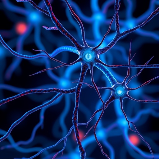
In a groundbreaking exploration into the intricate dialogue between nerves and cancer cells, recent research has unveiled a remarkable phenomenon: the direct transfer of mitochondria from peripheral nerves to cancer cells during metastasis. This discovery not only enriches our understanding of tumor microenvironment interactions but also opens new vistas for targeting cancer progression through neural modulation. The research, conducted using sophisticated imaging, genetic labeling, and sequencing techniques, highlights how neurons can actively contribute to cancer cell bioenergetics by donating their mitochondria, reshaping long-standing paradigms of cancer biology.
Human prostate cancer samples exhibiting perineural invasion—a condition where cancer cells encroach upon or surround nerves—displayed a significantly elevated mitochondrial load within cancer cells proximal to nerves. Using high-throughput multispectral imaging coupled with machine learning algorithms, researchers quantified mitochondrial abundance across tens of thousands of cells. Their precise spatial analysis revealed that cancer cells neighboring nerves harbored considerably more mitochondria than their distant counterparts, suggesting a nerve-dependent metabolic advantage in these cancer cells.
To further dissect whether this phenomenon involved actual mitochondrial transfer rather than mere mitochondrial biogenesis within cancer cells, the research team leveraged a clinical trial involving chemical denervation of the human prostate using botulinum neurotoxin A (BoNT/A). This intervention selectively reduced neuronal activity at the tumor site, serving as a natural experiment to observe the influence of neuronal innervation on cancer mitochondrial content. Remarkably, cancer cells on the denervated side exhibited a stark reduction in mitochondrial load, directly linking neuronal presence to mitochondrial enrichment in tumor cells and strongly implying transfer of mitochondria from nerves to cancer.
.adsslot_7eGRWNdP2H{width:728px !important;height:90px !important;}
@media(max-width:1199px){ .adsslot_7eGRWNdP2H{width:468px !important;height:60px !important;}
}
@media(max-width:767px){ .adsslot_7eGRWNdP2H{width:320px !important;height:50px !important;}
}
ADVERTISEMENT
To substantiate these findings with robust in vivo models, the researchers employed a BALB/c mouse xenograft system, where murine dorsal root ganglion (DRG) neurons innervating the mammary fat pad were genetically modified to express a mitochondria-targeted green fluorescent protein (GFP) reporter under the synapsin-1 promoter, allowing for neuron-specific mitochondrial labeling. Following the injection of 4T1 breast cancer cells tagged with a red fluorescent protein (mCherry) into the same fat pad, analysis of emerging tumors by flow cytometry detected a distinct subpopulation of cancer cells emitting the GFP mitochondrial signal. This provided compelling evidence that mouse host neurons transferred mitochondria directly into malignant cells within the tumor microenvironment.
Notably, this transfer was shown to be specific to mitochondria, as a control lentiviral construct encoding a nucleus-localized GFP failed to show any signal exchange, ruling out nonspecific protein transfer or artifact. This elegant genetic design confirmed that intact mitochondria, rather than soluble proteins or other organelles, crossed from host neurons into cancer cells in situ, emphasizing the precision and selectivity of this intercellular communication.
The implications of nerve-to-cancer mitochondrial transfer are profound, as mitochondria are central to energy production, metabolic adaptation, and apoptotic regulation. Mitochondrial acquisition by cancer cells could endow them with enhanced bioenergetic capacity, resistance to metabolic stress, and even influence metastatic potential. This discovery adds a new dimension to tumor–nerve interactions, previously focused largely on growth factor secretion and neurotransmitter release, by highlighting organelle-level communication.
From a therapeutic standpoint, these findings suggest new intervention points. Chemical denervation or strategies to block mitochondrial transfer mechanisms may disrupt this symbiotic relationship, potentially depriving cancer cells of vital metabolic support and hindering tumor growth and metastasis. Furthermore, the use of neuron-specific mitochondrial reporters in preclinical models paves the way for real-time imaging and therapeutic monitoring in future studies.
The researchers’ methodical approach, combining clinical patient sample analysis, advanced imaging platforms, genetic manipulation, and rigorous sequencing, exemplifies the power of multidisciplinary strategies in unraveling complex cellular communications. Their use of machine learning for image quantification allowed unprecedented cellular resolution and statistical power to detect subtle spatial mitochondrial variations linked to nervous system presence.
As cancer research continues to unravel the tumor microenvironment’s multifaceted influencers, the role of nerves emerges as a critical, active participant rather than a mere passive structure. The revelation that neurons can donate mitochondria to cancer cells during metastasis challenges classical tumor biology and invites new perspectives on tumor–neuron crosstalk.
Future research will need to clarify the precise molecular machinery mediating mitochondrial transfer, such as tunneling nanotubes, extracellular vesicles, or synaptic-like junctions, and how these processes are regulated by the tumor microenvironment. Understanding the downstream metabolic and signaling consequences of mitochondrial acquisition on cancer cell behavior will be equally critical.
In sum, this pioneering study illuminates a novel biological axis in cancer metastasis—the nerve-to-cancer mitochondrial conduit—adding an organelle-centric lens to investigate tumor progression and offering novel targets for therapeutic innovation. As interdisciplinary efforts converge, exploiting neural physiology to impede cancer’s metabolic support systems may forge new frontiers in oncology.
Subject of Research: Mitochondrial transfer from nerves to cancer cells during cancer metastasis
Article Title: Nerve-to-cancer transfer of mitochondria during cancer metastasis
Article References:
Hoover, G., Gilbert, S., Curley, O. et al. Nerve-to-cancer transfer of mitochondria during cancer metastasis. Nature (2025). https://doi.org/10.1038/s41586-025-09176-8
Tags: cancer cell metabolism and nerveshigh-throughput imaging in cancer researchimpact of botulinum neurotoxin on cancerinnovative cancer treatment strategiesmachine learning in cancer analysismitochondrial bioenergetics in tumorsmitochondrial transfer in cancernerves and cancer cell interactionneural modulation in cancer therapyprostate cancer perineural invasiontumor microenvironment and metastasisunderstanding cancer biology through nerve interactions

