In a groundbreaking advancement that promises to reshape the landscape of glioblastoma research and treatment, a team of researchers led by Yu, Basu, Baquer, and their colleagues have unveiled a novel investigative approach utilizing needle core biopsies to enable comprehensive multimodal deep-data generation. Their study, published in Nature Communications, volume 16, article 3957 (2025), introduces a powerful methodology that bridges the gap between clinical tissue sampling and high-dimensional molecular profiling, offering new hope for precision oncology in one of the most lethal brain cancers.
Glioblastoma, a highly aggressive and notoriously heterogeneous tumor of the central nervous system, has long posed formidable challenges to oncologists and neuroscientists alike. Its infiltrative growth patterns and rapid evolution thwart conventional treatments, resulting in dismal patient outcomes. Recognizing these challenges, the researchers focused on improving the quality and utility of biopsy samples, which historically have been limited by small tissue size and sampling bias. By employing investigative needle core biopsies, the team demonstrated the feasibility of acquiring robust, representative tumor samples suitable for extensive multimodal analysis.
Multimodal deep-data generation constitutes a convergence of various high-throughput and high-resolution technologies that collectively interrogate tumor biology at multiple layers, including genomic, transcriptomic, proteomic, and spatial contexts. The innovative sampling method elucidated by the study enables the extraction of precious tissue cores from live glioblastoma patients with minimal invasiveness, while preserving the architectural and molecular integrity essential for subsequent analyses. This technical refinement paves the way for integrative analyses that can decode the complex ecosystem of glioblastoma tumors.
Once the biopsy material was obtained, the team employed a suite of advanced multiplexed analyses. Single-cell RNA sequencing allowed for the dissection of individual tumor cells and surrounding microglia populations, revealing transcriptional states and identifying rare subpopulations potentially driving invasive behavior. Concurrently, spatial transcriptomics provided a map of gene expression distribution within the biopsied tissue architecture, uncovering spatial niches that might serve as therapeutic vulnerabilities or refuges from the immune system.
Complementing transcriptomic data, proteomic profiling was integrated through mass spectrometry techniques, capturing post-translational modifications and signaling network activities that are often decoupled from mRNA expression. This proteogenomic approach offered insights into the functional consequences of genetic alterations, helping to clarify how mutational landscapes translate into phenotypic traits that affect tumor aggressiveness and treatment resistance.
Additionally, advanced imaging modalities were incorporated, including multiplex immunohistochemistry and fluorescence in situ hybridization, to anatomically validate molecular data and preserve spatial context. These imaging strategies enabled the visualization of critical cell-cell interactions, vascularization patterns, and immune infiltration dynamics that collectively shape tumor behavior and response to therapies.
One of the pivotal outcomes of this research is establishing a standardized pipeline that converts limited biopsy material into comprehensive datasets amenable to machine learning and artificial intelligence analyses. Leveraging computational biology tools, the researchers created integrative models capable of predicting tumor evolution trajectories and patient-specific therapeutic responses. This big-data paradigm, rooted in reliable sample acquisition, heralds a move towards truly individualized medicine in glioblastoma care.
The implications of this work extend beyond glioblastoma itself. Many solid tumors share the challenge of heterogeneity and sampling limitations. Thus, the methodology and technological fusion proposed could be adapted for other malignancies, addressing a universal bottleneck in cancer biology—the ability to capture detailed, multidimensional data from clinically accessible tissue.
Furthermore, this approach opens avenues for longitudinal studies. By enabling repeat biopsies with minimal risk, clinicians can monitor tumor evolution and treatment response dynamically, rather than relying on static snapshots. This temporal dimension adds critical depth to precision oncology, allowing for adaptive therapeutic strategies that respond promptly to tumor adaptations.
Despite the promise, the study acknowledges inherent challenges. The integration of multimodal datasets demands advanced bioinformatics expertise and computational infrastructure, which may not yet be widely available in all clinical settings. Additionally, standardizing tissue handling and preserving sample integrity across multiple centers require collaborative efforts and rigorous quality controls to ensure data consistency and reproducibility.
Nevertheless, the potential benefits far outweigh these hurdles. The research team highlights that the combination of minimally invasive biopsy techniques with state-of-the-art analytical technologies effectively circumvents previous constraints, producing a foundation for discovering novel biomarkers, therapeutic targets, and mechanisms of resistance.
In conclusion, the investigative needle core biopsy approach is a transformative advancement in glioblastoma research, achieving a delicate balance between clinical practicality and scientific rigor. By unlocking the ability to generate deep, multimodal datasets from limited patient-derived tissue, it empowers researchers and clinicians with richer biological insights and steers the field towards more effective, personalized treatments.
As this methodology is adopted and refined, future studies will likely elucidate even more intricate dynamics within glioblastoma ecosystems and potentially identify strategies to overcome its stubborn therapeutic resistance. This research not only exemplifies innovation at the intersection of clinical practice and molecular science but also marks a hopeful turning point for patients afflicted by this devastating disease.
Subject of Research:
Glioblastoma needle core biopsies enabling multimodal molecular and imaging data generation.
Article Title:
Investigative needle core biopsies support multimodal deep-data generation in glioblastoma.
Article References:
Yu, K.K.H., Basu, S., Baquer, G. et al. Investigative needle core biopsies support multimodal deep-data generation in glioblastoma. Nat Commun 16, 3957 (2025). https://doi.org/10.1038/s41467-025-58452-8
Image Credits:
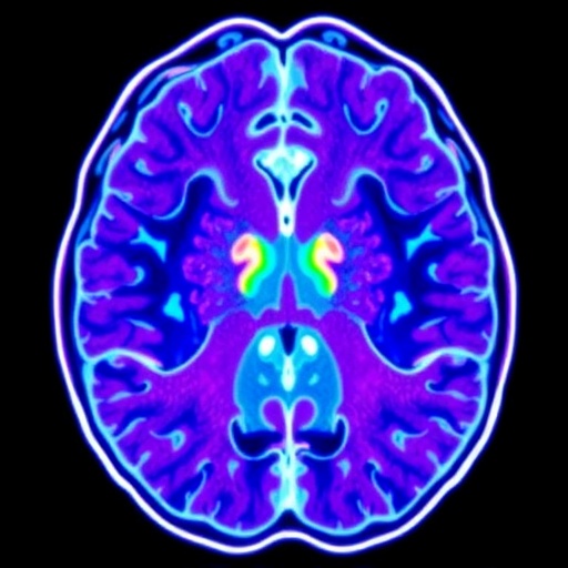
AI Generated
Tags: biopsy quality improvementchallenges in glioblastoma treatmentcomprehensive tumor samplingglioblastoma research advancementshigh-dimensional molecular profilinghigh-throughput biological technologiesinfiltration patterns of glioblastomamultimodal deep-data generationneedle core biopsiesneuro-oncology research innovationsprecision oncology techniquestumor biology analysis methods

