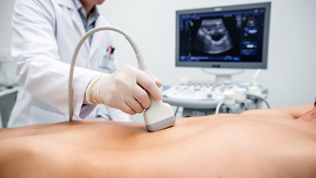
A narrative review published in Pain and Therapy synthesizes published anatomical and imaging literature to propose a standardized, surface landmark-based scanning algorithm for selected ultrasound-guided peripheral nerve blocks of the upper and lower limbs in orthopedic surgery. Ultrasound-guided regional anesthesia typically demands two cognitively distinct operational phases: image acquisition, wherein the clinician manipulates the transducer to identify the target neural and fascial anatomy, and image maintenance, wherein that optimized sonic view is actively preserved while advancing a block needle toward the designated structure. The authors observe that without a pre-formed mental expectation, ultrasound scanning frequently degrades into an unguided, open-ended search across the tissue plane. To address this challenge, the review formalizes a framework designed to structure image acquisition as an organized confirmatory sequence by systematically linking identifiable musculoskeletal surface landmarks to distinct sonographic endpoints through predetermined probe trajectories.
The conceptual framework divides procedural ultrasound into two separate cognitive burdens, each presenting unique psychomotor requirements for operators. Image acquisition demands sonoanatomical pattern recognition and precise transducer handling governed by hand-eye coordination, whereas image maintenance requires consistent alignment between the acoustic beam and the needle shaft alongside continuous, real-time visualization of the needle tip. Because an inadequate or suboptimal image at the end of the acquisition phase inevitably complicates subsequent needle guidance, instructional techniques established during initial scanning exert a direct influence over the entire nerve block procedure. The authors note that while reference atlases and digital materials widely describe isolated block targets, anatomical guidance remains fragmented across disparate regional descriptions rather than unified into an overarching scanning algorithm. Clinical society guidelines frequently outline training pathways and identify structures to recognize after scanning is underway, but they routinely omit the explicit surface landmarks from which ultrasound exploration should initially begin.
To bridge the operational gap between initial body inspection and final acoustic target recognition, the authors formulated a four-step sequence designated as Anchor, Place, Trace, and Confirm. Under this algorithm, the operator begins by establishing an Anchor, defined as a musculoskeletal surface landmark localized directly through visual inspection or light manual palpation without requiring measurement calipers or reliance on non-visible, deep structures. The second step, Place, derives an initial transducer position directly from that anchor, specifying the contact point and probe orientation relative to regional body axes. The third step, Trace, outlines an explicit search vector along which the probe is slid or rotated across the patient’s skin toward the target anatomy. Finally, the Confirm step defines the internal sonographic landmark that verifies correct transducer alignment over the therapeutic window, signaling the safe completion of image acquisition and the formal transition into the needle advancement phase.
Because the publication is a narrative synthesis of previously conducted investigations, it incorporates no prospective clinical trials, interventional protocols, or newly generated patient datasets. To illustrate the scanning sequences, the authors constructed illustrative figures pairing artificial intelligence-rendered surface anatomy diagrams with unmanipulated, newly acquired ultrasound images. The surface panels were generated from clinical photographs of a single healthy adult volunteer who was not a patient and from whom no diagnostic or personal medical data were gathered. Ultrasonic panels were obtained from this volunteer or, when acoustic resolution was insufficient for didactic publication, scanned directly from the review’s primary author. Both contributors provided formal written consent, and institutional ethics committee oversight was deemed unnecessary under Declaration of Helsinki principles because no diagnostic procedures or clinical interventions occurred. Every rendered anatomical panel was subsequently cross-checked against source photographs and evaluated for skeletal accuracy and limb posture by the corresponding author.
For upper limb regional anesthesia, the algorithm establishes two primary anchors that govern access to the cervical plexus and brachial plexus along a continuous neural pathway: the angle of the mandible and the apex of the deltopectoral groove. The mandibular angle, palpable at the junction of the mandibular ramus and body, anchors a cranial-to-caudal search vector along the sternocleidomastoid muscle. Placing the probe transversely over the sternocleidomastoid at this level identifies the great auricular nerve as a small hypoechoic oval on the anterior muscle surface. Sliding the transducer caudally traces this nerve until it disappears beneath the posterior border of the sternocleidomastoid at the punctum nervosum, where branches of the supraclavicular nerve and transverse cervical nerve emerge. Continuing the trace deeper through the prevertebral fascia leads directly into the interscalene groove, where the C5 and C6 nerve roots appear in cross-section between the anterior and middle scalene muscles.
The second upper limb anchor, the apex of the deltopectoral groove, marks the visible structural cleft between the deltoid and pectoralis major muscles at the junction of the lateral and middle thirds of the clavicle. By placing the probe immediately medial to this apex beneath the middle third of the clavicle, the clinician directly visualizes the costoclavicular space, where the lateral, posterior, and medial cords of the brachial plexus cluster in a consistent triangular arrangement lateral to the axillary artery. From this single stable reference point, pivoting the probe caudally around the apex into a parasagittal plane brings the distal infraclavicular cords into view at the coracoid process level. Alternatively, sliding the transducer cranially across the clavicle navigates directly into the supraclavicular fossa, where following the superior trunk as it divides into the C5 and C6 roots provides a cranial traceback to the interscalene level.
In the lower limb, regional anchors are arranged systematically across the inguinal region, thigh, and knee. Within the anterior groin, the anterior superior iliac spine and the pubic origin of the adductor longus serve as primary landmarks, defining an oblique geometric line that establishes starting positions for femoral nerve, fascia iliaca, iliopsoas plane, and pericapsular nerve group blocks. The authors highlight the adductor longus tendon origin as a superior medial reference compared to the traditional inguinal crease, noting that the medial aspect of the skin crease fades gradually into perineal soft tissue without a fixed skeletal boundary. For lateral coverage, the anterior superior iliac spine also anchors the lateral femoral cutaneous nerve block. Scanning transversely roughly ten centimeters distal to this spine enables clinicians to locate the nerve within the fat-filled flat tunnel formed by fascia lata duplicature between the sartorius and tensor fasciae latae muscles before tracing it cranially.
Secondary derived reference points further facilitate complex regional approaches in the pelvic and thigh regions. Positioning the transducer immediately lateral to the midpoint of the line connecting the anterior superior iliac spine to the adductor longus origin aligns the scan with the femoral head and indirect tendon of the rectus femoris, confirming the plane for femoral nerve, infrainguinal fascia iliaca, and iliopsoas plane blocks. Sliding the probe cranially along this fascial plane reveals the anterior inferior iliac spine and flat ilium for the pericapsular nerve group block, while revealing anisotropic signal changes as the femoral nerve curves over the pelvic brim. For obturator nerve blockade, placing the probe over the adductor longus origin and rotating toward the pectineus differentiates anterior and posterior branches or captures the common nerve trunk. Further distal, the midpoint of a line connecting the anterior superior iliac spine to the base of the patella guides transducer placement over the distal femoral triangle, where walking the probe caudally to the intersection of the sartorius and adductor longus muscle borders identifies the apex of the femoral triangle and the entrance to the adductor canal.
Around the knee joint, distinct tendinous anchors guide distal sciatic and terminal saphenous nerve procedures. At the posterolateral knee, the junction of the biceps femoris tendon and the popliteal crease anchors the popliteal sciatic nerve block. Transverse probe placement over this junction visualizes the common peroneal nerve and lateral sural cutaneous nerve superficial to the lateral head of the gastrocnemius muscle. Tracing these neural structures proximally reveals the tibial nerve and popliteal artery emerging from a deeper medial plane, serving as an intermediate waypoint before reaching the true sciatic bifurcation, where both major branches become enclosed within a single paraneural sheath. The authors note that in roughly twenty percent of individuals, the short head of the biceps femoris extends aberrantly distal, requiring clinicians who fail to locate the common peroneal nerve in superficial fat to seek it within a deeper intermuscular tunnel.
At the medial aspect of the knee, the pes anserinus tendon complex—representing the conjoined insertion of the sartorius, gracilis, and semitendinosus tendons along the anteromedial tibia—serves as the surface anchor for the saphenous nerve block. By placing the transducer transversely across this visible and palpable tendinous insertion and walking the probe caudally into the upper medial calf, operators can reliably locate the sartorial branch of the saphenous nerve, also known as the medial crural cutaneous nerve. Sonographically, this nerve presents as a small hyperechoic ovoid structure within the subcutaneous tissue plane situated superficial to the crural fascia. The great saphenous vein lies in the same superficial layer and assists in locating the tiny nerve structure, although operators must maintain light transducer pressure during scanning because excessive contact force can readily compress the fragile vein out of view.
The review emphasizes that the Trace step is not strictly mandatory for every procedure; when initial placement immediately generates an unambiguous acoustic endpoint, direct needle intervention may proceed without continuous displacement. Tracing is primarily advantageous when an isolated sonographic cross-section lacks defining features or when tracking a continuous neural structure confirms its anatomical identity. Furthermore, completing the Confirm step requires thorough visual interrogation of surrounding vascular and visceral structures rather than focusing narrowly on the target nerve alone. Operators must routinely identify adjacent large vascular channels such as the subclavian, axillary, femoral, and popliteal vessels, as well as the pleural line in thoracic-adjacent blocks. Additionally, clinicians should deliberately deploy color Doppler imaging to identify smaller, variable vessels that traverse needle paths, including the transverse cervical and dorsal scapular arteries in the neck, thoracoacromial branches in the infraclavicular fossa, and the superficial saphenous vein.
Crucially, the authors underline significant limitations surrounding their proposed framework, emphasizing that the central hypothesis—that anchor-based scanning reduces cognitive demand, shortens acquisition time, or enhances block success—remains an untested theoretical model requiring prospective clinical validation. No empirical measurements of operator cognitive load, procedure duration, or complication rates were collected or analyzed in this work. Furthermore, the physical reliability of surface anchors across diverse patient populations remains uncharacterized, particularly in patients presenting with morbid obesity, severe tissue edema, muscular atrophy, or traumatic skeletal deformities where surface contours and tactile bony landmarks become effaced. While structured rehearsal offers a standardized cognitive scaffold for intermediate practitioners and trainees, it cannot replace comprehensive anatomical mastery, and the algorithm does not claim to deliver demonstrated improvements in procedural safety or patient outcomes.
Subject of Research: Surface landmark scanning algorithm for ultrasound-guided orthopedic nerve blocks
Article Title: A Surface Landmark-Based Scanning Algorithm for Selected Ultrasound-Guided Nerve Blocks of the Upper and Lower Limb in Orthopedic Surgery: A Narrative Review
Article References: Yeoh, S.-R., Yang, M.-T., Wu, I. C., Horng, H.-C., Yang, S.-F., Chang, W.-W., & Lin, J.-A. (2026). A Surface Landmark-Based Scanning Algorithm for Selected Ultrasound-Guided Nerve Blocks of the Upper and Lower Limb in Orthopedic Surgery: A Narrative Review. Pain and Therapy. https://doi.org/10.1007/s40122-026-00903-1
Image Credits: AI Generated
DOI: 10.1007/s40122-026-00903-1
Keywords: ultrasound-guided nerve blocks, regional anesthesia, orthopedic surgery, surface landmarks, scanning algorithm, sonoanatomy, brachial plexus, cervical plexus, femoral nerve block, fascia iliaca, sciatic nerve block, adductor canal, pericapsular nerve group, pes anserinus, deltopectoral groove, image acquisition
News Source: Ophelia Keating. (November 16, 2026). Narrative Review Outlines Surface Landmark Scanning Framework for Orthopedic Nerve Blocks. Scienmag.