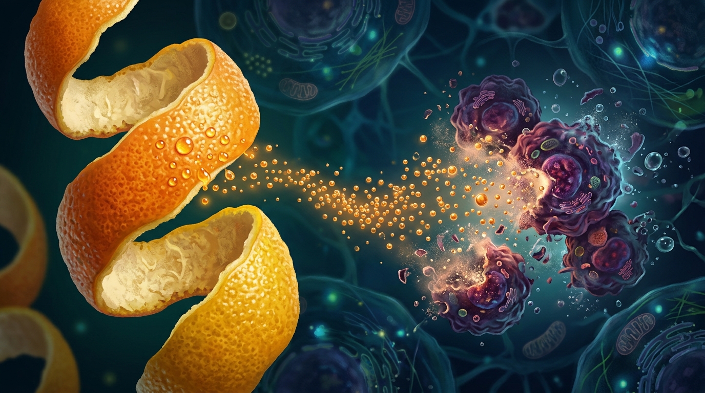
Nanoparticles prepared from the peel of Citrus unshiu Markovich, a citrus fruit cultivated on South Korea's Jeju Island, showed in vitro cytotoxic and apoptosis-inducing activity against HeLa cervical cancer cells in a study published in Frontiers in Nanotechnology. Researchers prepared an extract from the peel of Citrus unshiu Markovich, a seedless mandarin widely cultivated on South Korea’s Jeju Island, and converted it into nanoscale particles without adding any carrier or stabilizing agent. When tested against HeLa cervical cancer cells, the nanoparticle formulation triggered apoptotic cell death in 54.4 percent of treated cells after 24 hours, compared with 16.5 percent for the free, unformulated extract. The work, led by Meeta Gera of Jeju National University together with colleagues at Sher-e-Kashmir University of Agricultural Sciences and Technology of Jammu and Kangwon National University, characterized the nanoformulation and compared its physicochemical properties and in vitro anticancer activity with those of the free extract.
The choice of starting material is rooted in traditional medicine. In Korean practice, the dried peels of CUM, known as jin-pi, have long been used to treat asthma, appetite loss, and gastrointestinal disorders. The fruit itself is economically significant, accounting for more than 30 percent of Korea’s total fruit production. Earlier pharmacological studies have attributed anti-inflammatory, anti-allergic, anti-anxiety, anti-cancer, and anti-diabetic activities to the peels and their constituents, with hesperidin, narirutin, naringin, and nobiletin identified as the main flavonoids. The authors note a practical constraint, however: the extract's poor solubility and bioavailability mean that high dosages would be required for cancer treatment, and the study states that nanoformulations can modify delivery, stability, cellular uptake, and release characteristics, advantages that the authors say need to be experimentally verified for each formulation.
To prepare the extract, the team dried CUM peels under ambient conditions, pulverized them into a fine homogeneous powder, and extracted 10 grams of the powder in 100 milliliters of methanol using an ultrasonicator for one hour, followed by maceration for 24 hours. The filtrate was centrifuged at 12,000 rpm for 15 minutes, the solvent evaporated at 60 degrees Celsius under vacuum, and the remaining material freeze-dried to yield a dry extract stored at minus 20 degrees Celsius. For the nanoparticle version, 1 milliliter of the methanolic peel extract was added dropwise to 50 milliliters of distilled water at 0.2 milliliters per minute, stirred at 200 to 800 rpm for 30 minutes, and then sonicated at 30 kHz and 100 watts for two hours. The resulting pale yellow-orange solution was concentrated and freeze-dried into roughly 5 milligrams of orange-brown powder.
Characterization confirmed that the particles fell squarely in the nanometer range. Field-emission scanning electron microscopy and high-resolution transmission electron microscopy revealed predominantly spherical, well-dispersed, and uniform particles with an average diameter of approximately 40 to 60 nanometers. Dynamic light scattering corroborated this, yielding an average particle size of 42.6 nanometers. Ultraviolet-visible spectroscopy showed maximum absorption at 470 nanometers, and Fourier-transform infrared spectroscopy displayed strong peaks at 3,545, 2,938, 2,917, and 3,283 inverse centimeters, attributed to different phenolic compounds, with additional peaks at 3,423 and 1,647 inverse centimeters linked to the carbon-carbon stretching of naringin. The authors acknowledge that zeta potential and polydispersity index measurements were not performed, leaving surface charge and colloidal stability questions open for future work.
The chemical profile of the underlying extract proved potent. Total phenolic content reached 53.93 milligrams per gram in gallic acid equivalents, while total flavonoid content measured 7.96 micrograms per gram. Antioxidant performance was assessed with three complementary assays. In the DPPH radical-scavenging test, the extract achieved 98.60 percent scavenging at its highest tested concentration, and the ABTS assay reached 99.96 percent. The ferric reducing antioxidant power assay yielded 335.08 plus or minus 4.88 micromoles of ferrous sulfate per liter at the top concentration of 250 micrograms per liter. These measurements, taken across concentrations from 50 to 250 micrograms per liter, demonstrate strong in vitro antioxidant capacity, which the researchers associate with polyphenolic compounds capable of neutralizing free radicals through electron or hydrogen donation.
High-performance liquid chromatography with diode-array detection identified three prominent peaks in both the extract and the nanoparticles, tentatively aligned by retention time and spectral characteristics to naringin, hesperidin, and quercetin. The team then ran molecular docking simulations to explore how these three compounds might interact with four apoptosis-related receptors: TNFR1, TNFR2, FAS, and TRAIL. Hesperidin showed the most favorable predicted interactions with TNFR1 and TNFR2, forming four and two hydrogen bonds respectively with residues including Lys32, Val14, Cys15, Asn93, and Tyr61. Naringin formed a single hydrogen bond with alanine 143 on FAS but scored comparatively lower, while quercetin formed one hydrogen bond with arginine 62 on TRAIL. The authors are careful to state that docking alone cannot confirm receptor activation or establish a causal mechanism for the cellular effects; these computational findings suggest possible molecular interactions that warrant further experimental investigation.
The central biological experiments focused on HeLa cells, a cervical cancer cell line obtained from the Korean Cell Line Bank and cultured under standard conditions. Using the MTT assay, the researchers evaluated free CUM extract, the nanoparticle formulation, and the standard chemotherapy drug doxorubicin across concentrations from 0.0 to 100 micrograms per milliliter for the CUM preparations. The nanoparticles significantly inhibited HeLa cell viability compared with the other treatment groups, and 30 micrograms per milliliter was identified as the IC50 concentration under the tested experimental conditions. This dose-dependent cytotoxicity established the benchmark concentration used in the subsequent apoptosis and gene-expression analyses.
Flow cytometry using Annexin V-FITC and propidium iodide staining quantified the mode of cell death. After 24 hours of treatment at the optimal concentration, the nanoparticles induced apoptosis in 54.4 percent of HeLa cells, while the free extract induced apoptosis in 16.5 percent, a difference the authors interpret as a stronger apoptosis-inducing effect of the nanoformulation under the tested conditions. Necrotic cell populations did not differ significantly between the two groups, and the authors state that these findings suggest the nanoformulation may enhance the pro-apoptotic activity of the CUM extract in HeLa cells in vitro. Hoechst 33258 staining showed apoptotic morphological changes in the nuclear chromatin of treated cells, and the authors report that CUM NPs showed a noticeably greater cytotoxic effect on the cancer cells than free CUM and doxorubicin.
Quantitative reverse-transcription polymerase chain analysis then probed the molecular machinery behind the observed cell death. In nanoparticle-treated cells, the anti-apoptotic gene Bcl-2, the inflammatory cytokine IL-6, and the invasion-related gene MMP-9 were significantly downregulated, while the pro-apoptotic genes caspase-9, Bax, and the tumor suppressor p53 were significantly upregulated, all at p less than 0.05 compared with control groups. The IL-6 reduction reached a reported p value of 0.001485 and MMP-9 a p value of 0.001137. The authors link this expression pattern to the intrinsic pathway of apoptosis, in which mitochondrial release of cytochrome-c promotes apoptosome formation and caspase-9 activation, a cascade consistent with the shift from anti-apoptotic to pro-apoptotic signaling recorded here.
The study’s authors frame these results as a foundation rather than a finish line. All findings come from in vitro experiments on a single cancer cell line, and the docking results remain computational hypotheses. The physicochemical stability tests showed that particle size increased progressively over time, with more pronounced enlargement at pH 7.5 than under acidic conditions at pH 4.5 and 5.5, and the drug-release study found little release at pH 7.4 after 12 hours but almost 80 percent release at pH 5, which the authors interpret as pH-responsive release at lower pH levels. The researchers recommend further studies using additional cancer cell lines and in vivo models to establish broader biological relevance and therapeutic potential. For now, the findings are limited to in vitro experiments: the nanoformulated CUM peel preparation showed greater cytotoxicity and apoptosis induction than the free extract in HeLa cells, accompanied by a gene-expression pattern consistent with apoptosis, and the authors recommend further studies using additional cancer cell lines and in vivo models.
Subject of Research: In vitro anticancer activity of Citrus unshiu Markovich peel extract nanoparticles against HeLa cervical cancer cells
Article Title: In vitro evaluation of the anticancer activity of Citrus unshiu markovich extract and its nanoparticles in HeLa cells
Article References: Gera, M., Sharma, N., Kour, S., & Lee, S.-J. (2026). In vitro evaluation of the anticancer activity of Citrus unshiu markovich extract and its nanoparticles in HeLa cells. Frontiers in Nanotechnology, 8, Article 1997477. https://doi.org/10.3389/fnano.2026.1997477
Image Credits: AI Generated
DOI: 10.3389/fnano.2026.1997477
Keywords: Citrus unshiu Markovich, nanoparticles, HeLa cells, cervical cancer, apoptosis, phytochemicals, antioxidants, hesperidin, naringin, quercetin, nanoformulation, in vitro study
News Source: Nathaniel Bowman. (November 4, 2026). Nanoparticles Made From Korean Citrus Peel Kill HeLa Cancer Cells in Lab Tests. Scienmag.