A team of researchers has unveiled a sequencing-free, amplification-free strategy for mapping gene regulation directly in space, tackling key bottlenecks that have limited current spatial transcriptomics. Traditional methods rely on next-generation sequencing, demanding complex sample preparation and costly instrument pipelines, and they often provide only a partial view of how cells control gene expression after transcription.
The new approach targets post-transcriptional regulation in fresh tissues while preserving cellular geography. Instead of capturing and reading RNA by sequencing, the method uses a nanoneedle array to extract messenger RNAs (mRNAs), microRNAs (miRNAs), and N6-methyladenosine (m6A)-modified RNAs at the subcellular level. This design aims to quantify regulatory molecules without amplification steps that can distort abundance measurements.
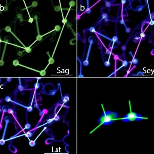
To read out what is collected, the researchers rely on multiplexed fluorescence encoding followed by imaging-based decoding. In effect, RNA targets are translated into fluorescence signatures that can be decoded computationally from microscopy, enabling spatially resolved profiles across large tissue areas. By operating on minimally processed fresh slices, the workflow avoids harsh fixation steps that can degrade sensitive RNA species.
Benchmarking against established standards strengthens the case for performance. The team compared their maps with fluorescence in situ hybridization, immunostaining, and bulk measurements. Reported results indicate sensitivity and spatial fidelity on par with conventional spatial transcriptomics approaches, while reducing both cost and operational complexity.
They also demonstrate biological utility by mapping patterned mRNA expression in developing mouse neural tissue. Such patterned readouts provide a stringent test of whether the system can faithfully reproduce structured expression landscapes rather than just detecting scattered signals.
As an even harder challenge, the method is applied to the olfactory bulb, a layered brain region known for pronounced spatial heterogeneity. Layered organization offers a rigorous scenario for evaluating whether regulatory profiles align with known anatomical structure.
Beyond model systems, the researchers extend the technology to human biopsy specimens. This compatibility suggests a path toward affordable spatial multi-omics-like analysis in clinical contexts, supporting disease stratification and prognostic assessment.
Overall, the study presents a streamlined route to spatially resolved post-transcriptional regulation that does not depend on sequencing. If broadly adopted, it could make high-resolution spatial biology more accessible—especially for fresh samples and laboratories where sequencing capacity is limited.
Subject of Research: Sequencing-free spatial profiling of post-transcriptional regulation in fresh tissues using nanoneedle arrays.
Article Title: Sequencing-free spatial profiling of post-transcriptional regulation in fresh tissues using nanoneedle arrays.
Article References: Ji, X., Fang, P., Wan, Y. et al. Sequencing-free spatial profiling of post-transcriptional regulation in fresh tissues using nanoneedle arrays. Nat. Biomed. Eng (2026). https://doi.org/10.1038/s41551-026-01745-0
DOI: https://doi.org/10.1038/s41551-026-01745-0
Keywords: Sequencing-free spatial transcriptomics; nanoneedle array; post-transcriptional regulation; microRNA; m6A; fluorescence encoding and imaging decoding; fresh tissues; subcellular resolution.
Tags: amplification-free molecular quantificationfresh tissue spatial profilingimaging-based RNA decodingmicroRNA and m6A detectionminimally processed tissue analysismultiplexed fluorescence imagingnanoneedle array gene mappingpost-transcriptional gene regulationsequencing-free gene expression profilingSpatial transcriptomicssubcellular RNA extractionTissue Architecture Preservation