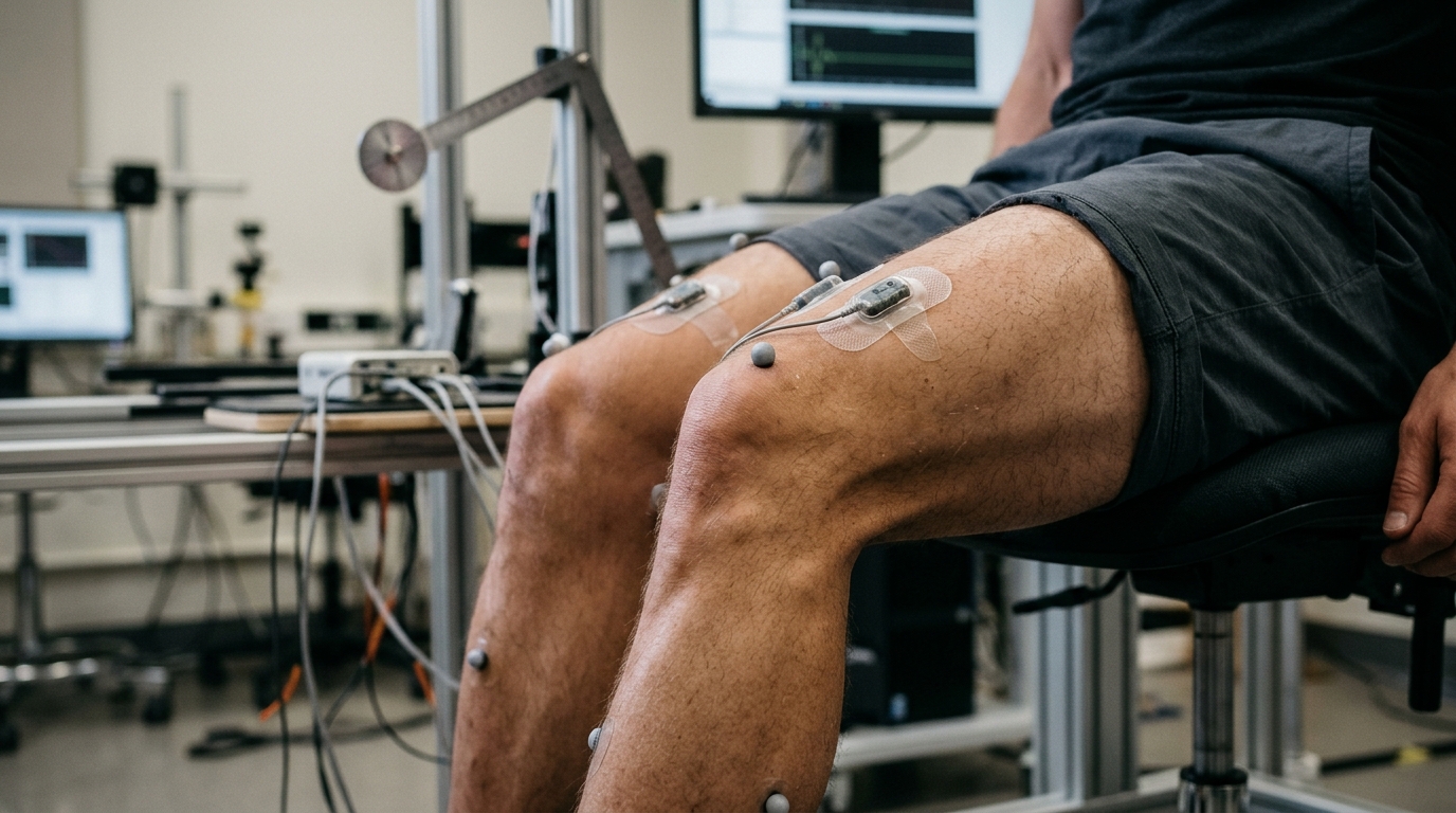
Measuring how stiff or supple a muscle is has long been a laboratory exercise performed on motionless limbs. Most established techniques for probing muscle viscoelasticity—the property that describes how muscle tissue combines spring-like elasticity with damping—work only when the joint is held still or moved very slowly. A new study published in the Journal of Medical and Biological Engineering proposes a way around that limitation, using a small sensor-based method called displacement mechanomyography to track muscle mechanical behavior while the knee is actually moving.
The research, led by Shinichi Fukuhara and Takaki Kawashima, set out to test whether the timing relationship between muscle deformation and joint motion could serve as a quantitative window into muscle viscoelastic properties. Their chosen tool, displacement mechanomyography or DMMG, records the mechanical deformation of the muscle itself, while joint kinematics capture how the limb is moving. If the muscle behaves like a viscoelastic material, its deformation should not perfectly coincide with the motion imposed on it; the lag between the two, expressed as a phase difference, might reveal how the tissue stores and dissipates mechanical energy in real time.
To test this idea, the team recruited twenty-five healthy young adults and divided them into a control group and a static stretching group. Static stretching served as a model intervention—a way of deliberately altering muscle mechanical state so the researchers could see whether their phase-based measurement would respond. During the experiment, DMMG signals were recorded from three muscles of the quadriceps group: the vastus medialis, the vastus lateralis, and the rectus femoris. Each participant’s leg was moved passively on a pedaling apparatus at two cadences, 10 and 50 revolutions per minute, meaning the muscles were stretched and shortened by the machine rather than by the participant’s own effort.
The core measurement was straightforward in concept. For each muscle and each condition, the researchers calculated the phase difference between the DMMG signal and the knee joint angle waveform. A small phase difference would suggest the muscle deforms nearly in step with the imposed motion, while a larger difference would indicate a greater lag—potentially reflecting different viscoelastic behavior.
The results showed a clear cadence effect in the control group. As pedaling speed increased from 10 to 50 rpm, phase differences between muscle deformation and knee angle rose markedly across all three recorded muscles. In other words, the faster the limb was moved, the more the muscle’s mechanical response lagged behind the joint’s motion. This cadence dependence is consistent with what viscoelastic theory would predict: tissues that combine elastic and viscous elements respond differently depending on the rate at which they are deformed.
The striking finding emerged when the researchers compared the two groups. In participants who had undergone static stretching, the cadence-dependent changes in phase difference were attenuated. At the faster 50 rpm cadence, phase differences were significantly smaller in the stretching group than in the control group. The stretching intervention appeared to have altered how the muscles responded mechanically during rapid passive movement, and the DMMG-based phase measurement detected that change.
Interestingly, the analysis found no significant differences in phase measurements between the three individual quadriceps muscles within the conditions tested. The effect of stretching showed up as a group-level difference at the higher cadence rather than as a redistribution of behavior among the vastus medialis, vastus lateralis, and rectus femoris.
The authors interpret these findings as evidence that DMMG–kinematic phase differences are sensitive to stretching-induced changes in muscle mechanical behavior during passive movement. Because the method requires only recording muscle deformation and joint angle during ordinary passive motion, it could in principle offer a non-invasive, muscle-specific, and objective approach to evaluating dynamic muscle mechanical behavior—something existing static or quasi-static techniques cannot provide.
The researchers are careful about what the study does and does not establish. The work was conducted in healthy young adults using a passive pedaling model, and the conclusion is framed as warranting further validation: phase-based DMMG analysis is presented as a promising candidate indicator of muscle viscoelasticity, not yet a validated clinical measure. Static stretching served as a model intervention to demonstrate sensitivity, and the study does not claim the method diagnoses any condition or predicts injury. What it does show is that a timing-based signal, measurable during movement, shifts in a systematic way when muscle mechanical state is altered—a result that could open the door to assessing muscle viscoelasticity in dynamic, real-world conditions rather than only on the examining table.
Subject of Research: Using displacement mechanomyography to quantify muscle viscoelastic properties during passive movement before and after static stretching
Article Title: Phase-Based Assessment of Muscle Viscoelastic Properties During Passive Movement using Displacement Mechanomyography following Static Stretching
Article References: Fukuhara, S., & Kawashima, T. (2026). Phase-Based Assessment of Muscle Viscoelastic Properties During Passive Movement using Displacement Mechanomyography following Static Stretching. Journal of Medical and Biological Engineering. https://doi.org/10.1007/s40846-026-01062-6
Image Credits: AI Generated
DOI: 10.1007/s40846-026-01062-6
Keywords: displacement mechanomyography, muscle viscoelasticity, static stretching, passive movement, phase difference, joint kinematics, quadriceps, biomechanics, DMMG, knee joint angle, muscle mechanical behavior, Phase-Based
News Source: Ophelia Keating. (November 6, 2026). Muscle Stiffness Tracked in Motion as Mechanomyography Signals Lag Behind the Knee. Scienmag.