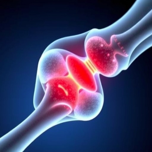
Bone fractures may be repaired with help from an unexpected source: dormant cells hiding inside skeletal muscle. A study published online in Bone Research has identified a population of muscle-resident progenitor cells that can leave their normal surroundings after injury, travel to a fracture, and transform into bone-forming cells. In mice, these cells generated a substantial portion of the osteoblasts responsible for rebuilding damaged bone and also contributed to the restoration of the bone marrow environment. The discovery challenges the traditional view that fracture repair depends mainly on cells already located within bone or its surrounding periosteum.
Bone healing is a highly coordinated biological process involving inflammation, blood-vessel formation, cartilage production, bone deposition, and tissue remodeling. Osteoblasts, the specialized cells that produce new bone matrix, are central to this process, but their origins can vary depending on the type and severity of injury. Researchers have increasingly recognized that cells outside the skeleton can remain in a quiet, undifferentiated state and acquire new functions when tissues are damaged. The new study, led by Dr. Ugur M. Ayturk and colleagues from the Skeletal Health and Orthopedic Research Program in the United States, investigated whether muscle-resident cells could participate directly in fracture repair.
The researchers focused on fibroadipogenic progenitors, commonly known as FAPs. These cells are found between muscle fibers and are normally involved in maintaining and repairing skeletal muscle. Despite their regenerative potential, FAPs can also contribute to scar formation or abnormal tissue growth under certain conditions. To follow their behavior, the team identified expression of the gene Clec3b as a highly specific marker of a normally dormant progenitor population. Using genetically engineered mice, the scientists attached a fluorescent label to cells expressing Clec3b, allowing them to track the cells before and after bone injury and determine what types of cells they eventually became.
Under normal growth conditions, the labeled cells remained in skeletal muscle and in the superficial layer of the periosteum, the thin connective-tissue membrane covering the outer surface of bone. They did not spontaneously enter the bone or develop into osteoblasts. Fracture changed this behavior dramatically. Following injury, Clec3b-labeled cells rapidly moved toward the damaged region and accumulated in the healing callus, the temporary tissue that forms around a fracture. As repair progressed, many of these cells stopped expressing Clec3b and acquired the characteristics of osteoblasts. This loss of the marker suggested that Clec3b identifies the cells in their dormant state rather than their mature, bone-forming state.
The contribution of these muscle-derived cells was unexpectedly large. Approximately three weeks after fracture, about 28 percent of the osteoblasts in the healing callus were traced back to Clec3b-lineage cells. These cells produced new bone matrix and became integrated into the developing repair tissue. Some also differentiated into bone marrow stromal cells, which do not directly form most of the mineralized bone but create the supportive cellular environment required inside the marrow cavity. By five weeks after injury, the descendants of the original dormant progenitors were therefore contributing not only to the hard outer structure of regenerated bone but also to the internal tissue that supports blood formation and bone maintenance.
The investigators used single-cell RNA sequencing to examine the molecular changes occurring during this transformation. This technique measures gene activity in individual cells and can reveal transitional states that are difficult to detect using conventional microscopy. The analysis showed that dormant Clec3b-lineage cells adopted gene-expression profiles associated with osteoblasts and bone marrow stromal cells after fracture. In other words, the cells did not simply migrate into the injury site; they underwent a defined biological reprogramming process. Their molecular identity shifted from a quiescent muscle-associated progenitor state toward distinct skeletal cell populations capable of rebuilding different components of the damaged bone.
Additional experiments indicated that skeletal muscle was the principal source of these regenerative cells. Similar Clec3b-positive cells were found in the superficial periosteum, raising the possibility that the periosteum might supply most of the progenitors during healing. However, when researchers surgically removed the periosteum before inducing fractures, labeled cells from muscle still traveled to the injury site and generated bone-forming descendants. Bone graft experiments provided further evidence: grafts that retained surrounding muscle produced considerably more Clec3b-lineage osteoblasts than grafts without muscle. These findings suggest that muscle adjacent to an injured bone is not merely a passive tissue surrounding the fracture but an active reservoir of cells with skeletal regenerative potential.
The same progenitor population also appeared to drive heterotopic ossification, a condition in which cartilage and bone form abnormally within muscles or other soft tissues after trauma, surgery, neurological injury, or severe burns. In mouse models, Clec3b-lineage cells developed into both cartilage-producing and bone-producing cells, becoming a major source of the unwanted mineralized tissue. When the researchers blocked a key pathway required for bone formation or depleted the Clec3b-positive cells, both normal fracture healing and heterotopic ossification were reduced. This dual role makes the cells biologically significant and clinically complicated: stimulating them could help difficult fractures heal, while suppressing them could potentially prevent disabling abnormal bone growth.
The findings point toward a new therapeutic strategy based on controlling the behavior of dormant extraskeletal progenitors rather than introducing external stem cells. Treatments that activate Clec3b-lineage cells, encourage their migration, or guide their differentiation could one day improve healing in patients with large bone defects, delayed unions, or impaired regenerative capacity. Conversely, drugs that interrupt the same pathways could help limit heterotopic ossification after major injuries. However, the research was conducted in mice, and the precise equivalents of these cells in humans remain to be established. Human studies will also need to determine how age, disease, inflammation, muscle damage, and the size of a fracture influence the cells’ behavior. Even with these limitations, the study reveals that skeletal muscle contains a previously underappreciated reserve of bone-forming potential—and that the body’s response to a fracture may begin far beyond the edge of the broken bone.
Subject of Research: Animals
Article Title: Clec3b+ extraskeletal cells regulate fracture healing and heterotopic ossification
News Publication Date: 6 July 2026
Web References: https://www.nature.com/boneres/
References: DOI: 10.1038/s41413-026-00532-6
Image Credits: Dr. Ugur M. Ayturk, Skeletal Health and Orthopedic Research Program
Keywords: bone healing, fracture repair, skeletal muscle, fibroadipogenic progenitors, FAPs, Clec3b, osteoblasts, bone marrow stromal cells, heterotopic ossification, regenerative medicine, tissue engineering, stem cells, orthopedics
Tags: bone fracture repair mechanismsbone marrow environment restorationbone tissue remodeling processescartilage production in fracture repaircellular sources of osteoblasts in bone repairchallenges to traditional fracture healing viewsinflammation and blood-vessel formation in healingmuscle-bone interaction in tissue regenerationmuscle-resident progenitor cellsosteoblast origin in fracture healingrole of dormant muscle cells in bone regenerationskeletal muscle contribution to bone healing