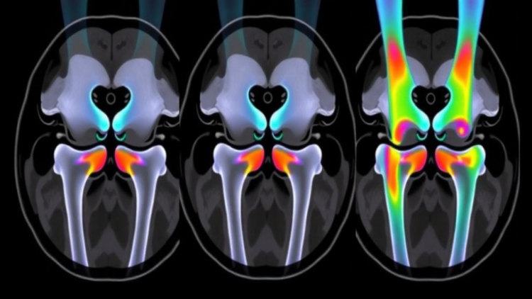
The knee of a child is not simply a smaller version of an adult knee. It is a structure in motion, with ligaments lengthening, bony grooves widening and growth plates slowly sealing shut in a sequence that differs between boys and girls. A new study published in BMC Medical Imaging has now charted that developmental choreography in detail, using magnetic resonance imaging to follow hundreds of children and adolescents between the ages of six and eighteen. The work, led by Yuepeng Qi and colleagues at Hebei Medical University Third Hospital in Shijiazhuang, China, offers one of the most systematic MRI-based portraits to date of how the anterior cruciate ligament, the intercondylar notch and the distal femur mature together, and it introduces a simple measurement that may help clinicians judge skeletal maturity with greater precision.
The anterior cruciate ligament, or ACL, is the strap of connective tissue that keeps the thigh bone from sliding forward over the shin bone. In adults, tears of this ligament are among the most common and consequential sports injuries, and in skeletally immature patients they pose a particular surgical dilemma: standard reconstruction techniques can damage the very growth plates that still have work to do. Understanding what a normal, developing ACL looks like on MRI at every age is therefore not an academic exercise. It is the reference frame against which abnormal anatomy, injury and surgical planning must be judged. Until now, the researchers note, age- and sex-related developmental patterns of the pediatric knee have remained incompletely characterized on routine imaging.
To fill that gap, the team conducted a retrospective single-center study of 520 children and adolescents aged six to eighteen years who had undergone knee MRI. From those scans, the investigators extracted a set of quantitative features: the length of the ACL, the width of the intercondylar notch, the height of the medial femoral epiphysis, abbreviated MEH, and the status of the distal femoral physis, the cartilaginous growth plate near the knee end of the thigh bone. The physis was assessed categorically, reflecting the familiar radiological progression from an entirely open plate, through partial closing, to a fully closed and fused structure. Statistical treatment of the data relied on generalized additive models, which allow flexible, non-linear relationships between age and anatomy to emerge, along with Spearman correlation, Welch analysis of variance and exploratory receiver operating characteristic analyses.
The results trace two parallel but offset developmental timelines. ACL length and intercondylar notch width both increased with age, but the curves flattened earlier in females than in males, a pattern consistent with the earlier onset and completion of skeletal maturation in girls. In other words, by the time boys are still adding length to their ligaments and width to the notch that cradles them, many girls have already reached their adult configuration. This sexual dimorphism matters for anyone interpreting a pediatric knee scan, because a measurement that is perfectly normal for a fourteen-year-old girl might signal delayed development in a boy of the same age. The generalized additive models captured these trajectories as continuous curves rather than abrupt stages, giving clinicians a smoother and more nuanced picture of normal variation.
The distal femoral physis told a complementary story. Its status progressed with age in the expected direction, from open toward closing and closed, and females transitioned earlier toward the closing and closed states. That finding will surprise no pediatric orthopedist, but its value lies in the quantification: the study ties the categorical physeal stages to a large, age-stratified MRI cohort, providing empirical grounding for the visual judgments radiologists make every day. Because the distal femoral physis contributes substantially to leg-length growth, its status is a critical variable in decisions ranging from ligament reconstruction timing to limb-lengthening procedures and the treatment of growth disturbances.
Perhaps the most intriguing contribution of the study is the medial epiphyseal height. This measurement, taken from the medial side of the distal femoral epiphysis, increased steadily with age and, crucially, was significantly associated with physeal status across the entire cohort and separately within each sex. Where the traditional physeal assessment sorts patients into discrete buckets, MEH offers a continuous, quantitative readout of local distal femoral development. The distinction is more than semantic. Continuous measures can track change within a stage, detect subtle deviations from the expected trajectory and potentially refine risk stratification in ways that categorical staging cannot. The authors propose that MEH may complement, rather than replace, the categorical assessment of the distal femoral physis.
The exploratory receiver operating characteristic analysis quantified just how informative that continuous measure might be. An ROC curve plots sensitivity against specificity across all possible cutoffs of a diagnostic test, and the area under that curve, the AUC, summarizes discriminative power on a scale from 0.5, equivalent to a coin flip, to 1.0, perfect classification. For distinguishing physeal status, MEH yielded an AUC of 0.855 in the overall cohort, rising to 0.923 in males and standing at 0.855 in females. Values in this range indicate good to excellent discrimination, suggesting that a single linear measurement already available on routine knee MRI carries substantial information about how far along a child is in the process of skeletal maturation.
Why does this matter beyond the radiology reading room? ACL injuries in children and adolescents are rising as youth sports participation intensifies, and surgeons increasingly face the question of how to reconstruct a torn ligament without injuring a growth plate that still has years of contribution to make to final leg length. Physeal-sparing techniques exist precisely for the skeletally immature, but choosing among them requires an accurate estimate of remaining growth. A continuous, MRI-derived marker such as MEH, correlated with physeal status and characterized across age and sex, could give surgical teams a finer-grained instrument for that estimation, complementing the traditional categorical staging that has long been the standard of care.
The study also carries implications for research into bone development more broadly. The authors frame their findings as a basis for future investigation in skeletally immature patients with ACL injury, and the age- and sex-specific developmental curves they report could serve as normative references in studies of growth disorders, juvenile idiopathic arthritis and other conditions that alter musculoskeletal maturation. Because the measurements come from routine MRI rather than specialized imaging or ionizing radiation, they could in principle be extracted from scans already obtained for clinical reasons, opening the door to large-scale, low-burden normative databases. The researchers acknowledge the exploratory nature of the ROC component, and the retrospective, single-center design means the findings will need validation in independent and more diverse cohorts before they change practice.
Methodological rigor receives attention as well. The team evaluated measurement reliability using intraclass correlation coefficients, a standard gauge of how consistently different observers or repeated measurements produce the same values, an essential step for any quantitative metric hoping to enter clinical use. Ethical oversight came from the Institutional Review Board of Hebei Medical University Third Hospital, which approved the retrospective design with a waiver of written informed consent, and the study was conducted according to the ethical standards of the 1964 Declaration of Helsinki. The work was supported by funding from Hebei Province and China’s National Key Research and Development Program. For now, the study’s legacy may be conceptual as much as practical: it demonstrates that the growing knee can be measured, not merely looked at, and that a single epiphyseal height on an ordinary MRI scan can speak volumes about the hidden timetable of skeletal maturity.
Subject of Research: Age- and sex-related MRI development of the pediatric knee, including ACL morphology, intercondylar notch shape and distal femoral physeal maturation
Article Title: Age- and sex-related MRI developmental patterns of the anterior cruciate ligament, intercondylar notch, and distal femur in children and adolescents
Article References: Age- and sex-related MRI developmental patterns of the anterior cruciate ligament, intercondylar notch, and distal femur in children and adolescents. (n.d.). https://doi.org/10.1186/s12880-026-02876-1
Image Credits: AI Generated
DOI: 10.1186/s12880-026-02876-1
Keywords: anterior cruciate ligament, pediatric knee, MRI, medial epiphyseal height, distal femoral physis, skeletal maturity, intercondylar notch, bone development, sexual dimorphism, musculoskeletal development, growth plate, ACL injury
Cite Scienmag News
APA MLA Chicago
Ophelia Keating. (October 1, 2026). MRI Maps How the Child Knee Grows: ACL, Notch and Growth Plate Development Tracked by Age and Sex. Scienmag. https://scienmag.com/mri-maps-how-the-child-knee-grows-acl-notch-and-growth-plate-development-tracked-by-age-and-sex/
Ophelia Keating. “MRI Maps How the Child Knee Grows: ACL, Notch and Growth Plate Development Tracked by Age and Sex.” Scienmag, 1 October 2026, https://scienmag.com/mri-maps-how-the-child-knee-grows-acl-notch-and-growth-plate-development-tracked-by-age-and-sex/. Accessed 1 October 2026.
Ophelia Keating. “MRI Maps How the Child Knee Grows: ACL, Notch and Growth Plate Development Tracked by Age and Sex.” Scienmag. October 1, 2026. https://scienmag.com/mri-maps-how-the-child-knee-grows-acl-notch-and-growth-plate-development-tracked-by-age-and-sex/
Copy citation Download RIS
Tags: ACL injuryACL maturation in childrenage-related changes in pediatric knee structuresanterior cruciate ligamentanterior cruciate ligament development in youthbone developmentChild knee developmentclinical implications of MRI in pediatric orthopedicsdistal femoral physisfemur growth and segmentationgrowth plategrowth plate development in adolescentsintercondylar notchintercondylar notch growth in childrenmedial epiphyseal heightMRIMRI imaging of pediatric knee growthMRI-based assessment of skeletal maturitymusculoskeletal developmentpediatric kneepediatric knee anatomy and biomechanicssex differences in knee growthsexual dimorphismskeletal maturity