Credit: Image created by M Röhrich, University Hospital Heidelberg, Germany.
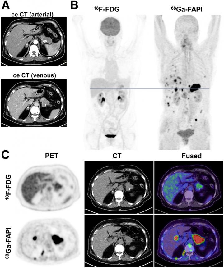
Reston, VA–For patients with pancreatic ductal adenocarcinomas (PDAC), molecular imaging can improve staging and clinical management of the disease, according to research published in the June issue of The Journal of Nuclear Medicine. In a retrospective study of PDAC patients, the addition of PET/CT imaging with 68Ga-FAPI led to restaging of disease in more than half of the patients, most notably in those with local recurrence.
PDAC is a highly lethal cancer, with a five-year survival rate of less than 10 percent. Optimal imaging of PDAC is crucial for accurate initial TNM (tumor, node, metastases) staging and selection of the primary treatment. Follow-up imaging is also important to accurately detect local recurrence or metastatic spread as early and as completely as possible.
“Currently, contrast-enhanced CT is the gold standard when it comes to TNM staging, and PET imaging isn’t typically part of the clinical routine” stated Manuel Röhrich, MD, nuclear medicine physician at Heidelberg University Hospital in Heidelberg, Germany. “However, we know that PDAC is composed of certain fibroblasts that express fibroblast activation protein, which can be imaged with the novel PET radiotracer 68Ga-FAPI. Given this characteristic, we sought to explore the utility of 68Ga-FAPI PET/CT to image FDAC patients.”
The study included 19 FDAC patients who received contrast-enhanced CT imaging followed by 68Ga-FAPI PET/CT. Results from the 68Ga-FAPI PET/CT scans were then compared with TNM staging based on contrast-enhanced CT. Changes in oncological management were recorded.
68Ga-FAPI PET/CT-based TNM staging differed from contrast-enhanced CT imaging in 10 out of 19 patients, which resulted in changes in TNM staging. Of the 12 patients with recurrent disease, eight were upstaged, one was downstaged and three remained the same. In the seven patients newly diagnosed with PDAC, one was upstaged, while the staging remained the same for six of the patients.
“This analysis suggests that 68Ga-FAPI PET/CT is a promising new imaging modality in staging of PDAC that may help to detect new or clarify inconclusive results obtained by standard CT imaging,” said Röhrich. He added, “Improvement in survival can only be achieved by effective treatment approaches customized to the individual patient’s disease status. Thus, hybrid imaging using FAPI tracer may open up new applications in staging and restaging of PDAC.”
###
The authors of “Impact of 68Ga-FAPI-PET/CT imaging on the therapeutic management of primary and recurrent pancreatic ductal adenocarcinomas” include Manuel Röhrich, Fabian Staudinger, Dawn, P. Liew, Clemens Kratochwil and Hendrik Rathke, Department of Nuclear Medicine, Heidelberg University Hospital, Heidelberg, Germany; Patrick Naumann, Jakob Liermann, Klaus Herfarth and Stefan A. Koerber, Department of Radiation Oncology, Heidelberg University Hospital, Heidelberg, Germany, National Center for Tumor diseases (NCT), Heidelberg, Germany, and Heidelberg Institute of Radiation Oncology (HIRO), Heidelberg, Germany; Frederik L. Giesel, Department of Nuclear Medicine, Heidelberg University Hospital, Heidelberg, Germany, and German Cancer Consortium (DKTK), partner site Heidelberg, Germany; Peter L. Choyke, Molecular Imaging Program, Center for Cancer Research, National Cancer Institute, National Institutes of Health, Bethesda, Maryland; Annika Wefers, Department of Neuropathology, Institute of Pathology, University Hospital Heidelberg, Heidelberg, Germany, and Clinical Cooperation Unit Neuropathology, German Consortium for Translational Cancer Research (DKTK), German Cancer Research Center (DKFZ), Heidelberg, Germany; Dirk Jäger, Department of Medical Oncology and Internal Medicine Virgin Islands, National Center for Tumor Diseases, University Hospital Heidelberg, Germany, and Clinical Cooperation Unit Applied Tumor Immunity, German Cancer Research Center (DKFZ), Heidelberg, Germany; Jürgen Debus, Department of Radiation Oncology, Heidelberg University Hospital, Heidelberg, Germany, National Center for Tumor diseases (NCT), Heidelberg, Germany, and Heidelberg Institute of Radiation Oncology (HIRO), Heidelberg, Germany, German Cancer Consortium (DKTK), Partner Site Heidelberg, Germany, Heidelberg Ion-Beam Therapy Center (HIT), Department of Radiation Oncology, Heidelberg University Hospital, Heidelberg, Germany, and Clinical Cooperation Unit Radiation Oncology, German Cancer Research Center (DKFZ), Heidelberg, Germany; Uwe Haberkorn, Department of Nuclear Medicine, Heidelberg University Hospital, Heidelberg, Germany, and German Cancer Consortium (DKTK), Partner Site Heidelberg, Germany, Clinical Cooperation Unit, Department of Nuclear Medicine, German Cancer Research Center (DKFZ), Heidelberg, Germany, and Translational Lung Research Center Heidelberg, Member of the German Center for Lung Research DZL, Heidelberg, Germany; and Matthias Lang, Department of Neuropathology, Institute of Pathology, University Hospital Heidelberg, Heidelberg, Germany, and Department of Surgery, Heidelberg University Hospital, Heidelberg, Germany, Member of the German Center for Lung Research DZL, Heidelberg, Germany.
This work was funded by the Federal Ministry of Education and Research, grant number 13N 13341. Uwe Haberkorn, Clemens Kratochwil and Frederik Giesel have filed a patent application for quinoline-based FAP-targeting agents for imaging and therapy in nuclear medicine. This retrospective study was approved by the local institutional review board (study number S115/2020).
Visit JNM‘s new website for the latest research, and follow our new Twitter and Facebook pages @JournalofNucMed.
Please visit the SNMMI Media Center for more information about molecular imaging and precision imaging. To schedule an interview with the researchers, please contact Rebecca Maxey at (703) 652-6772 or [email protected]
About JNM and the Society of Nuclear Medicine and Molecular Imaging
The Journal of Nuclear Medicine (JNM) is the world’s leading nuclear medicine, molecular imaging and theranostics journal, accessed more than 11 million times each year by practitioners around the globe, providing them with the information they need to advance this rapidly expanding field. Current and past issues of The Journal of Nuclear Medicine can be found online at http://jnm.
JNM is published by the Society of Nuclear Medicine and Molecular Imaging (SNMMI), an international scientific and medical organization dedicated to advancing nuclear medicine and molecular imaging–precision medicine that allows diagnosis and treatment to be tailored to individual patients in order to achieve the best possible outcomes. For more information, visit http://www.
Media Contact
Rebecca Maxey
[email protected]
Original Source
http://www.
Related Journal Article
http://dx.


