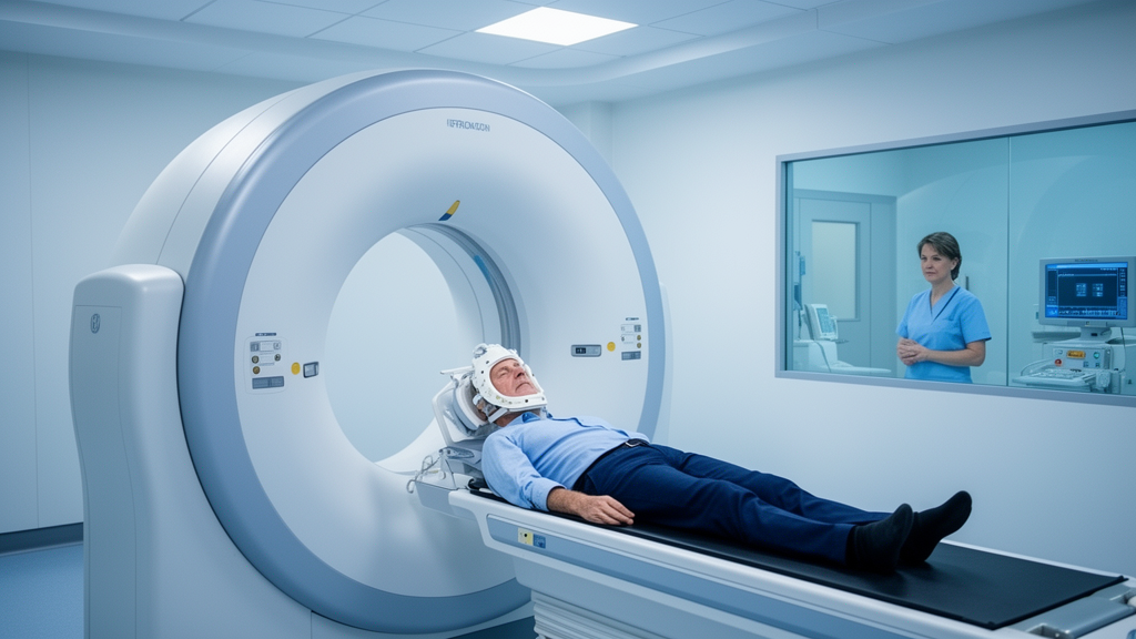
For the tens of thousands of people treated each year for cancers of the throat, tonsil and base of tongue, the radiation beam that destroys the tumor often claims an unintended casualty: the salivary glands. Sitting just millimeters from the treatment targets, these glands are exquisitely sensitive to radiation, and their damage leaves many survivors with a parched, fragile mouth that complicates eating, speaking and dental health for years. A new clinical study from North Macedonia now offers some of the most detailed objective evidence yet that newer radiation technology can meaningfully blunt that damage.
…
The biological basis for this vulnerability lies in the architecture of the salivary glands themselves. The serous acinar cells that produce the watery component of saliva are among the most radiosensitive cell populations in the human body, and they are largely replaced by fibrotic, fatty tissue when radiation doses exceed the tolerance of the glandular parenchyma. Within the first week of a conventional fractionated course, salivary output can fall by half or more, and the saliva that remains is often thick, viscous and acidic, with reduced buffering capacity and altered electrolyte concentrations. These biochemical shifts help explain why post-radiation patients face a cascade of oral problems beyond dryness itself: accelerated dental caries that characteristically attacks the cervical margins of the teeth, difficulty wearing dentures, mucosal soreness, and heightened risk of candidal infections.
The distinction between the two techniques compared in the study rests on how each shapes the radiation dose distribution. Three-dimensional conformal radiotherapy, the older of the two approaches, shapes beams to match the outline of the tumor as seen from several fixed directions, but the intensity of each beam is essentially uniform across its width. Volumetric modulated arc therapy, by contrast, rotates the treatment machine continuously around the patient while the beam intensity is modulated by a moving multileaf collimator and the dose rate is varied in real time. This allows the high-dose region to be sculpted with far greater precision around irregular targets while deliberately steering low-dose corridors away from organs at risk, including the parotid and submandibular glands. The result is a dose distribution that can spare substantial volumes of glandular tissue that would otherwise receive damaging levels of radiation.
The magnitude of the differences reported is striking. Patients treated with 3DCRT had a median unstimulated whole-saliva flow of 0.07 mL per minute, a value that falls well below the commonly used threshold for hyposalivation, whereas the VMAT group’s median of 0.14 mL per minute, while still reduced compared with healthy norms, represents a doubling of resting secretion. Stimulated whole-saliva flow showed an even larger relative gap, with medians of 0.25 and 0.50 mL per minute respectively. The parotid-specific measurements are particularly informative, because the parotid glands contribute the bulk of stimulated saliva and are known to be highly sensitive to mean radiation dose. A median stimulated parotid flow of 0.24 mL per minute in the VMAT group, more than twice the 0.10 mL per minute observed after 3DCRT, is consistent with the idea that the modulated technique preserved more functional acinar tissue in these glands.
The modified Schirmer test adds a complementary dimension to the assessment. Originally developed in ophthalmology to quantify tear production, the test has been adapted for oral use by placing standardized filter paper strips against the oral mucosa and measuring the wetted length in millimeters. It captures moisture at the mucosal surface, which reflects not only salivary output but also the practical wetting environment that oral tissues actually experience. The median wetted length of 13 millimeters in the VMAT group versus 7 millimeters in the 3DCRT group suggests that the functional advantage of the newer technique extended beyond laboratory flow measurements to the clinical reality of mucosal hydration.
The measurement protocol itself deserves attention, because salivary flow rates are notoriously sensitive to collection conditions. Unstimulated whole saliva, collected by the spitting method, represents the basal secretory output of all the major and minor glands combined and is the standard measure for diagnosing hyposalivation. The stimulated collections in this study used two different approaches: electrical stimulation with a SaliPen device, which delivers a mild electrical stimulus to evoke reflex secretion, and gustatory stimulation with modified Lashley cups positioned over the parotid ducts, which isolates the output of the parotid glands specifically. Separating unstimulated and stimulated measurements, and distinguishing whole-mouth from gland-specific output, allows a more nuanced picture of which parts of the salivary system were affected, since different glands have different dose-response relationships and different contributions to oral comfort under resting versus eating conditions.
The three-month time point is also meaningful in the natural history of radiation injury. Salivary dysfunction after radiotherapy follows a biphasic pattern, with an acute inflammatory phase during and shortly after treatment followed by a chronic phase in which progressive fibrosis, vascular damage and acinar cell loss dominate. By three months, the acute phase has largely subsided, so the differences observed at this interval likely reflect a mixture of residual acute recovery and emerging chronic damage. Some recovery of salivary function can continue for a year or more after treatment, particularly in glands that received lower doses, which means the gap between the two techniques could either narrow or widen with longer follow-up. Longitudinal data would be needed to determine which trajectory these cohorts follow.
Several caveats temper the interpretation of the findings. The allocation between techniques was not randomized; radiation oncologists selected the modality they considered most appropriate for each individual patient, which raises the possibility that the groups differed in tumor extent, location, or other characteristics that independently influence salivary outcomes. Tumors situated closer to the parotid glands, for example, may have been more likely to receive one technique over the other, and the required radiation doses may have differed between groups. The cross-sectional design, in which patients were assessed at a single time point after treatment rather than followed prospectively from a pre-treatment baseline, means that pre-existing differences in salivary function cannot be excluded. The authors appropriately acknowledge these limitations, noting that causality cannot be established from this design.
It is also worth noting that all patients in both groups received concurrent weekly cisplatin, the standard radiosensitizing chemotherapy for oropharyngeal cancer. Chemotherapy can itself contribute to mucosal toxicity and may compound the oral sequelae of radiation, but because both groups received the same regimen, this factor is unlikely to explain the observed differences. The uniformity of the chemotherapy protocol is in fact a methodological strength, isolating the radiotherapy technique as the principal variable between the groups.
The clinical stakes of these findings are considerable. Xerostomia consistently ranks among the most burdensome late effects reported by head and neck cancer survivors, affecting quality of life measures as strongly as many tumor-related symptoms. No pharmacological therapy reliably restores radiation-damaged salivary tissue; management currently relies on sialagogues such as pilocarpine, artificial salivas, meticulous dental care and dietary modification, all of which provide partial relief at best. Prevention through dose-sparing treatment planning therefore remains the most effective strategy, and the adoption of intensity-modulated techniques has been driven in large part by the goal of reducing parotid dose. The present study contributes objective, multi-parameter evidence to that effort, measuring not just subjective dryness but quantifiable secretion rates from different glands and mucosal hydration directly.
For patients and clinicians, the practical message is that the choice of radiation delivery technique can have measurable consequences for oral function months after treatment ends. As VMAT becomes increasingly available in radiation oncology centers worldwide, studies like this one, which document its effects on organ function rather than only on tumor control, help inform shared decisions about treatment planning. Future research with prospective designs, pre-treatment baseline measurements, longer follow-up and larger cohorts will be needed to confirm whether the salivary advantages observed here persist over years and translate into durable improvements in quality of life for head and neck cancer survivors.
Subject of Research: Salivary gland function after 3DCRT versus VMAT radiotherapy for oropharyngeal cancer
Article Title: Assessment of salivary gland function after head and neck radiotherapy using 3DCRT and VMAT: a comparative clinical study
Article References: Assessment of salivary gland function after head and neck radiotherapy using 3DCRT and VMAT: a comparative clinical study. (n.d.). https://doi.org/10.3389/froh.2026.1934444
Image Credits: AI Generated
DOI: 10.3389/froh.2026.1934444
Keywords: radiotherapy, VMAT, 3DCRT, salivary gland function, xerostomia, head and neck cancer, oropharyngeal cancer, salivary flow, Schirmer test, parotid gland, cisplatin, radiation oncology
News Source: Nathaniel Bowman. (October 16, 2026). Modern Arc Radiotherapy Keeps More Saliva Flowing After Head and Neck Cancer Treatment. Scienmag.