


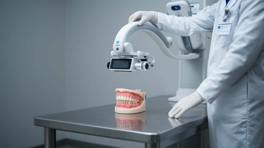
Forensic teams trying to identify disaster victims could soon have a new imaging tool small enough to wheel into a temporary mortuary. A study published in the Egyptian Journal of Forensic Sciences reports that a modern mobile 3D C-arm cone-beam computed tomography system completed postmortem dental imaging in 15 forensic cases without a single technical interruption, producing volumetric datasets that captured teeth, metallic dental findings, and root canal fillings while generating panoramic-like images and 3D reconstructions from one scan.
The research, led by Dominic Gascho and published on 20 August 2026, set out to test whether a compact intraoperative imaging platform could generate the dental datasets that disaster victim identification, or DVI, depends on. Forensic odontology remains one of the primary identifiers recognized by INTERPOL, and dental structures are highly resistant to decomposition, thermal damage, and fragmentation, making them particularly valuable when other identifying features are compromised. Radiological documentation of dental morphology, restorations, implants, and endodontic treatments is central to comparing antemortem and postmortem records, yet dedicated dental imaging remains difficult in temporary mortuary facilities, remote locations, and disaster environments.
The system under evaluation was the Cios Spin, a mobile 3D C-arm CBCT unit made by Siemens Healthineers, which the manufacturer provided for a two-week evaluation period in May 2025. Over 10 working days, the team imaged postmortem cases that were already undergoing routine postmortem imaging, adding the CBCT examination only when it did not interfere with the routine forensic workflow. All examinations used the manufacturer’s standard 3D acquisition protocol for obese patients: a 30-second rotational acquisition with 400 projections over an angular range of approximately 195 degrees, at 111 kV and 100 mA with 0.1 mm copper filtration. The reconstructed datasets contained isotropic voxels of 0.313 mm in an 800 by 800 by 512 matrix, corresponding to an image volume of roughly 25 by 25 by 16 centimeters.
From that single acquisition, the researchers generated multiplanar reformations, maximum intensity projections, curved planar reconstructions that produced panoramic-like images of both dental arches, and 3D volume renderings. The study cohort comprised nine males and six females with a median age of 60 years, ranging from 22 to 91. Five individuals had a postmortem interval of less than 4 days, while the remaining ten had estimated intervals ranging from approximately 1 to 2 weeks up to 8 to 24 weeks. The dentition was visualized in all 13 dentate cases; the other two were completely edentulous. Metallic dental findings were present in 13 of 15 cases and endodontic treatments in four, and where present, these features were identifiable on the reconstructed images.
Technical feasibility was assessed qualitatively by a single imaging scientist with 19 years of experience in diagnostic radiology and 14 years in postmortem CT imaging. The assessment criteria included image acquisition without technical interruption, complete volumetric coverage of the maxilla and mandible, visualization of teeth and identification-relevant findings, and successful generation of all reconstruction formats. Coverage of the maxilla and mandible was achieved in all 15 cases without repeat acquisition, despite variable head positioning and different stages of postmortem change. The term metallic dental findings was used as a descriptive imaging term for radiopaque metallic dental structures visible on the images, with no further classification of type or number performed.
One of the more technically interesting results involved reconstruction kernels and metal artefact reduction. Reconstruction using the blur-reducing kernel appeared to improve the delineation of fine dental structures compared with the standard kernel, particularly in regions containing metallic findings. The manufacturer’s metal artefact reduction algorithm reduced streak artefacts in adjacent anatomical regions, but it did not improve visualization of the metallic dental findings themselves and in some cases altered their appearance. The authors note this is consistent with previous forensic imaging studies showing that such algorithms reduce streak artefacts caused by metallic objects without necessarily improving visualization of the objects. For detailed evaluation of metallic findings, appropriate reconstruction and display parameters remain essential.
Display settings turned out to matter as much as artefact reduction. The Cios Spin reconstructs volumetric datasets with a 16-bit depth, enabling extended windowing and improved visualization of high-density structures. Metallic dental findings were best visualized using dedicated high-density window settings within the CT-compatible DICOM viewer, with displayed window values of approximately 5,000 HU centre and 15,000 HU width, values the authors stress served solely for image visualization and do not represent quantitatively validated Hounsfield units, an inherent limitation of cone-beam CT technology. Occasionally, metallic implants exhibited higher radiodensity than other metallic restorations and required even wider display windows for optimal visualization, under which the contours and morphology of the findings could be seen more clearly than with standard settings.
For comparison, all 15 cases underwent high-resolution fan-beam CT immediately before the CBCT examination during the same imaging session, on a SOMATOM Definition Flash scanner using a dedicated dental protocol with 120 kV, 300 mAs, pitch 0.75, 0.4 mm slice thickness, and an Ur68u reconstruction kernel, reconstructed with the extended CT scale. In qualitative side-by-side review, the mobile 3D C-arm datasets appeared to show reduced blooming artefacts associated with metallic dental findings compared with the fan-beam images, so that root canal filling material and adjacent dental structures remained more clearly delineated in the presented examples. The authors emphasize that this comparison was illustrative only, was not performed under standardized experimental conditions, and permits no conclusions about comparative diagnostic performance between the modalities.
The study’s limitations are explicit. It was a prospective exploratory technical feasibility study with a limited number of cases, selected to assess feasibility and therefore not representative of the full spectrum of forensic odontological casework. Image evaluation was performed by a single experienced observer without assessment of interobserver agreement, so the observations represent a qualitative assessment rather than a formal evaluation of diagnostic accuracy or observer performance. No antemortem to postmortem image matching or deployment under operational disaster victim identification conditions was performed, meaning the study demonstrates technical feasibility only and does not allow conclusions about diagnostic performance or effectiveness within real DVI workflows. Future studies should incorporate standardized image quality metrics, multiple independent observers, and objective performance measures.
Even so, the operational implications are striking. Mobile multidetector CT systems have already been deployed during large-scale DVI operations, including the 2017 Grenfell Tower disaster, where on-site postmortem CT combined with remote image interpretation facilitated timely radiological documentation and reporting. After the 2011 Great East Japan Earthquake and tsunami, by contrast, CT imaging could not be implemented during the identification process despite the scale of the incident, mainly because of logistical constraints, distributed mortuary sites, and the limited deployability of conventional CT systems. Unlike conventional multidetector CT installations, mobile 3D C-arm CBCT systems require comparatively little infrastructure and can be operated using standard electrical power supplies, which may facilitate deployment in temporary mortuary facilities, remote locations, field operations, or disaster response scenarios where installing a full-size CT scanner is logistically challenging. The authors position the system not as a replacement for multidetector CT but as a complementary modality that may extend access to dedicated postmortem dental imaging where conventional infrastructure is unavailable. They also point out that the standardized volumetric datasets could support future artificial intelligence-assisted forensic odontology, an area of rapidly expanding research that remains investigational. Further studies are warranted to evaluate diagnostic performance and operational implementation in DVI settings.
Subject of Research: Feasibility of mobile 3D C-arm cone-beam CT for postmortem dental imaging in forensic odontology and disaster victim identification
Article Title: Postmortem dental imaging using a mobile 3D c-arm cone-beam CT system: a feasibility study for forensic odontology and disaster victim identification
Article References: Gascho, D. (2026). Postmortem dental imaging using a mobile 3D c-arm cone-beam CT system: a feasibility study for forensic odontology and disaster victim identification. Egyptian Journal of Forensic Sciences, 16(1), Article 75. https://doi.org/10.1186/s41935-026-00575-0
Image Credits: AI Generated
DOI: 10.1186/s41935-026-00575-0
Keywords: forensic odontology, disaster victim identification, cone-beam CT, postmortem imaging, mobile C-arm, dental radiography, metal artefact reduction, panoramic reconstruction, forensic radiology, temporary mortuary, 3D imaging, technical feasibility
News Source: Ophelia Keating. (October 30, 2026). Mobile 3D C-arm cone-beam CT shows feasibility for postmortem dental imaging in disaster victim identification. Scienmag.



