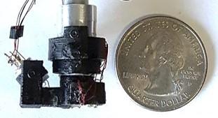
Credit: Arvind Pathak
Working with mice, a team of Johns Hopkins Medicine researchers has developed a relatively inexpensive, portable mini microscope that could improve scientists’ ability to image the effects of cancer, stroke, Alzheimer’s disease and other conditions in the brains of living and active mice over time. The device, which measures less than 5 cubic centimeters, is docked onto animals’ heads and gathers real-time images from the active brains of mice moving naturally around their environments.
“This technology allows us to record really rich amounts of data on the underlying functions of the brain over the lifetime of a disease model,” says Arvind Pathak, Ph.D., associate professor of radiology and biomedical engineering at the Johns Hopkins University School of Medicine, and a member of the Johns Hopkins Kimmel Cancer Center.
A report on the development of the mini microscope was published Jan. 9 in Nature Communications.
Traditional microscopes used in brain imaging studies are large stationary microscopes that can be prohibitively expensive and can cost tens of thousands of dollars, say researchers, limiting the number of labs capable of imaging over long periods of time.
Additionally, the bulky nature of benchtop microscopes requires laboratory animals to be completely still for imaging. This often requires animals be repeatedly anesthetized to get clear images. Anesthetized brains undergo changes unrelated to disease, potentially muddying the waters between real results and the brain’s response to the anesthetic drug.
The new microscope, which functions like a mini GoPro action camera, is able to capture images in real-time and is fully portable. This eliminates the need to anesthetize animals for imaging, allowing researchers to observe disease changes in a more natural state and relate such changes to the animal’s behavior.
In contrast to other mini microscopes, the new microscope offers researchers three imaging options to observe changes in the mouse brain as a disease progresses over time: fluorescence imaging to observe neurons firing or track fluorescently tagged cells; so-called intrinsic optical signal imaging to observe changes in the structure of blood vessels across the course of a disease; and laser speckle contrast imaging to follow changes in blood flow as a disease progresses.
Pathak and his team built the prototype device using commercially available miniature components, such as LED lights, microscope lenses, image sensors and custom-made 3D printed components. The housing, which docks the microscope onto a mouse’s head, is entirely 3D printed and reusable. The whole setup, lead author Janaka Senarathna says, then plugs into a laptop computer where researchers can collect and analyze the images.
In a proof-of-concept experiment designed to follow the course of a brain tumor, the research team injected the brains of mice with human brain cancer cells genetically engineered to glow so they can be seen by the microscope. They then mounted the microscope onto the mouse’s head, and continuously imaged the mice over 16 days.
In the images gathered during this time, the researchers were able to watch new blood vessels grow alongside the tumor as the cancer progressed. The researchers were then able to measure the blood flow changes during the dynamic remodeling of these blood vessels.
“We successfully monitored these microscopic changes on a daily basis, which allowed us to watch aspects of the disease in remarkable detail,” says Pathak.
One remarkable aspect, Pathak said, is that this microscope could be a powerful tool for imaging the effect of new drugs for such diseases.
“This is just one example of the utility of this technology, and one that could someday have an impact on how best to assess response to treatments,” says Pathak.
The researchers, who are working with Johns Hopkins Technology Ventures to spin off this technology, say a commercial version might cost approximately 10 times less than currently available models, and they plan to refine the device to capture clearer images and monitor additional brain functions.
###
Other researchers involved in this study include, Hang Yu, Callie Deng, Alice Zou, John Issa, Darian Hadjiabadi, Stacy Gil, Qihong Wang, Betty Tyler and Nitish Thakor of the Johns Hopkins University School of Medicine.
This research was supported by the National Cancer Institute (1R21CA175784-01, 1R01CA196701, P30NS050274) and a Kavli neuroscience distinguished fellowship.
Janaka Senarathna, Hang Yu, Nitish Thakor and Arvind Pathak have an international patent (PCT/US18/40979) pending. The remaining authors declare no competing interests.
Media Contact
Rachel Butch
[email protected]
Original Source
https:/

