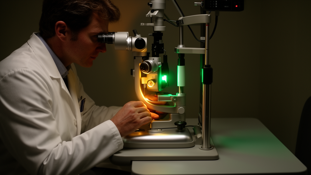
In a prospective, randomized controlled trial conducted at Affiliated Jinhua Hospital of Zhejiang University School of Medicine, researchers have demonstrated that 360-degree MicroPulse laser trabeculoplasty provides superior intraocular pressure lowering compared with guideline-escalated medical therapy in patients with diverse open-angle glaucoma subtypes. The study, which evaluated 90 adult participants over a six-month duration, revealed that laser intervention achieved a 30.6 percent reduction in intraocular pressure alongside a 61.1 percent drop in daily medication burden. Furthermore, advanced anterior segment imaging showed quantifiable anatomical expansion within the conventional aqueous humor drainage pathway, providing rare randomized clinical evidence linking procedural pressure reduction to structural changes within Schlemm’s canal.
Open-angle glaucoma represents the predominant cause of irreversible blindness worldwide, accounting for an estimated 70 to 80 percent of all glaucoma diagnoses and imposing substantial burdens on healthcare infrastructure. The underlying pathophysiology centers on impaired aqueous humor outflow through the trabecular meshwork and Schlemm’s canal, which causes intraocular pressure to rise. Elevated intraocular pressure remains the sole clinically modifiable risk factor proven to retard progressive optic neuropathy and subsequent visual field loss.
To investigate a non-incisional alternative that avoids the thermal coagulative damage typical of older laser modalities, the trial protocol compared single-session circumferential MicroPulse laser application against standardized medical escalation. Between February and April 2026, investigators screened 142 individuals, ultimately enrolling 90 patients who were randomly allocated in an equal ratio to either the laser or medication group. Eligible participants were 18 years or older presenting with primary open-angle glaucoma, normal-tension glaucoma, ocular hypertension, or secondary open-angle glaucoma. Individuals displaying angle closure, active intraocular inflammation, previous laser or filtration procedures, or significant corneal disease affecting imaging quality were systematically excluded.
Patients allocated to the laser cohort received treatment using a 577-nanometer yellow laser platform configured to deliver 1000 milliwatts of power with a 15 percent duty cycle, consisting of 0.5-millisecond on-time and 2.83-millisecond off-time bursts across a 300-micrometer spot size. A total of 120 spots, divided evenly at 30 applications per quadrant, were directed to the pigmented trabecular meshwork at the Schlemm’s canal junction under direct gonioscopic visualization. Because all participants were already using topical pressure-lowering drops at baseline with an average of 1.8 medications per patient, baseline pharmaceutical regimens in the laser cohort were intentionally tapered and discontinued over the initial postoperative month, being reinstated only if intraocular pressure exceeded individual target levels.
In contrast, participants in the control group followed guideline-escalated medical protocols based on regional and European standards. Medical management commenced with once-daily prostaglandin analogs, followed by sequential addition of topical beta-blockers or carbonic anhydrase inhibitors if pressure targets, defined as a 20 to 30 percent reduction from baseline or a severity-adjusted safe threshold, were not reached. Allocation concealment was maintained through an independent centralized random sequence generator, and while operating physicians could not be masked to procedural execution, all clinical technicians performing automated tonometry, corneal specular microscopy, visual field testing, and optical coherence tomography analysis were rigorously masked to study assignment throughout the trial.
Primary efficacy assessments revealed marked differences in intraocular pressure trajectories between the cohorts. At baseline, mean intraocular pressure measured 24.2 millimeters of mercury in the laser cohort and 23.9 millimeters of mercury in the medical group. By month six, pressure in the laser group dropped to a mean of 16.8 millimeters of mercury, reflecting an absolute reduction of 7.4 millimeters of mercury, or 30.6 percent. The medical control group reached a mean pressure of 18.7 millimeters of mercury, corresponding to a reduction of 5.2 millimeters of mercury, or 21.8 percent. A linear mixed-effects model with repeated measures established a statistically significant between-group difference of 2.2 millimeters of mercury favoring the laser arm, with responder analysis indicating that 82.2 percent of laser-treated patients achieved at least a 20 percent pressure drop compared with 55.6 percent in the control arm.
High-resolution anterior segment optical coherence tomography yielded significant mechanistic observations regarding Schlemm’s canal morphology. Baseline canal diameters were comparable between cohorts, measuring 287.4 micrometers in the laser arm and 291.2 micrometers in the control arm. By month three, the laser cohort exhibited an expansion of 37.2 micrometers to reach 324.6 micrometers, while the medication group showed a non-significant variation of 2.3 micrometers to 293.5 micrometers, resulting in a statistically significant adjusted between-group difference of 34.9 micrometers. Concurrently, angle opening distance and trabecular-iris space area expanded significantly following laser intervention. Statistical analysis revealed a moderate positive correlation between month-three canal expansion and intraocular pressure reduction within the laser cohort, supporting biological hypotheses that subthreshold stimulation promotes structural remodeling of outflow channels.
Corneal safety and visual stability remained intact across both treatment arms over six months of clinical observation. Central endothelial cell density evaluations via specular microscopy confirmed that laser administration caused no structural injury to the corneal endothelium, with cell counts declining by an insignificant 1.5 percent in the laser group and 1.4 percent in the medication arm. Hexagonal cell ratios and coefficients of variation showed equivalent stability. Visual functional measures, including standard automated perimetry mean deviation, and structural parameters such as retinal nerve fiber layer thickness and ganglion cell complex dimensions exhibited no adverse deterioration or significant between-group divergence, demonstrating that pressure-lowering interventions effectively maintained optic nerve preservation.
Secondary clinical outcomes documented substantial advantages for laser intervention regarding quality of life and procedural safety. Patient-reported Glaucoma Quality of Life-15 questionnaire scores demonstrated a 5.8-point improvement in the laser cohort compared to a 1.1-point change in controls. Postoperative laser complications were mild and self-limited, consisting of transient pressure elevation in three patients that resolved spontaneously by day three and mild anterior chamber inflammation in three patients that resolved within a week under standard topical care. In contrast, 26.7 percent of control patients developed medication-related adverse events requiring clinical intervention, predominantly ocular surface toxicity and systemic beta-blocker effects.
The study authors emphasize several methodological limitations that warrant consideration when interpreting these findings. The single-center setting and six-month follow-up timeframe limit generalizability and preclude evaluation of long-term disease progression or permanent visual field preservation beyond the primary endpoint. Furthermore, participants could not be masked to procedural versus topical therapies, which may have introduced expectancy effects on patient-reported quality of life metrics. Exploratory completer subgroup analyses indicated that secondary open-angle glaucoma patients showed the largest pressure reduction of 9.1 millimeters of mercury, yet small subgroup sample sizes make these findings hypothesis-generating. Future multi-center investigations with extended surveillance and head-to-head comparisons against selective laser trabeculoplasty will be essential to define the long-term durability of MicroPulse therapy.
The temporal trajectory of intraocular pressure reduction underscored notable mechanistic differences between the two treatment approaches. Analysis using a linear mixed-effects model identified a significant group-by-time interaction, reflecting a rapid therapeutic response in the laser arm that materialized by week one, achieving a mean reduction of 4.6 millimeters of mercury compared to 1.8 millimeters of mercury with topical therapy, and remained stable through month six. Beyond the twenty percent responder threshold, nearly half of the laser-treated participants (48.9 percent) achieved an intraocular pressure reduction of at least thirty percent at six months, compared with only 20.0 percent in the medication cohort. These findings proved resilient to participant attrition; among the 78 patients who completed the six-month protocol out of 90 originally randomized, a complete-case sensitivity analysis confirmed an identical between-group difference of 2.2 millimeters of mercury favoring laser trabeculoplasty.
Stratified analyses across specific open-angle glaucoma subtypes offered further granular insights into clinical responsiveness. Among completers diagnosed with primary open-angle glaucoma, laser therapy produced a mean pressure drop of 7.8 millimeters of mercury compared with 5.4 millimeters of mercury under medical management. Participants with normal-tension glaucoma exhibited a smaller yet statistically significant reduction of 3.2 millimeters of mercury versus 1.8 millimeters of mercury in medically managed controls. When evaluating the anatomical mechanism, researchers noted that the observed correlation between Schlemm’s canal expansion and pressure lowering (Pearson r = 0.62) does not definitively prove direct causation, as higher baseline intraocular pressure could independently allow greater structural distension and broader scope for pressure reduction.
At the cellular level, the biological effects of subthreshold micropulse irradiation are thought to stem from non-destructive photostimulation that triggers local cytokine release and extracellular matrix turnover within the trabecular apparatus. The investigators emphasized that maintaining pristine structural anatomy leaves conventional filtration procedures and surgical options fully available should patients experience disease progression later in life.
Subject of Research: Evaluation of 360-degree MicroPulse laser trabeculoplasty versus stepped medical therapy in open-angle glaucoma
Article Title: 360° Micropulse laser trabeculoplasty versus medical therapy in open-angle glaucoma: a randomized trial of efficacy, safety, and anterior segment morphological changes
Article References: Xie, P., He, N., Li, X., He, J., & Bao, J. (2026). 360° Micropulse laser trabeculoplasty versus medical therapy in open-angle glaucoma: a randomized trial of efficacy, safety, and anterior segment morphological changes. Frontiers in Medicine, 13, Article 1985905. https://doi.org/10.3389/fmed.2026.1985905
Image Credits: AI Generated
DOI: 10.3389/fmed.2026.1985905
Keywords: glaucoma, open-angle glaucoma, micropulse laser trabeculoplasty, intraocular pressure, schlemm's canal, ophthalmology, anterior segment OCT, clinical trial, eye drops, aqueous outflow, trabecular meshwork, randomized controlled trial, corneal endothelium, glaucoma quality of life, laser surgery, ocular hypertension, vision care
News Source: Ophelia Keating. (November 17, 2026). MicroPulse Laser Outperforms Medication in Open-Angle Glaucoma Trial. Scienmag.