Electroconvulsive therapy triggers widespread yet spatially convergent structural and functional alterations across the brain in patients treated for psychiatric disorders, according to a systematic review and voxel-based meta-analysis published in Translational Psychiatry.
Researchers conducted the investigation to address historically inconsistent findings across individual neuroimaging studies regarding electroconvulsive therapy. While the intervention is established as an effective treatment across multiple mental disorders, previous structural and functional imaging reports had identified conflicting regional patterns across patient cohorts.
To locate spatially convergent effects, the team screened literature across PubMed, Web of Science, and Embase. The literature search identified studies evaluating patients with mental disorders before and after electroconvulsive therapy using voxel-based morphometry to assess brain structure alongside resting-state voxel-based pathophysiology to evaluate brain function.
The meta-analysis integrated data from 45 eligible studies encompassing 55 individual experiments and a pooled cohort of 1395 psychiatric patients. Across this synthesized dataset, the researchers carried out 23 distinct meta-analyses using the activation likelihood estimation algorithm to measure pre- and post-treatment changes.
Analytical comparisons evaluated datasets independently by imaging modality as well as pooled across multimodal datasets.
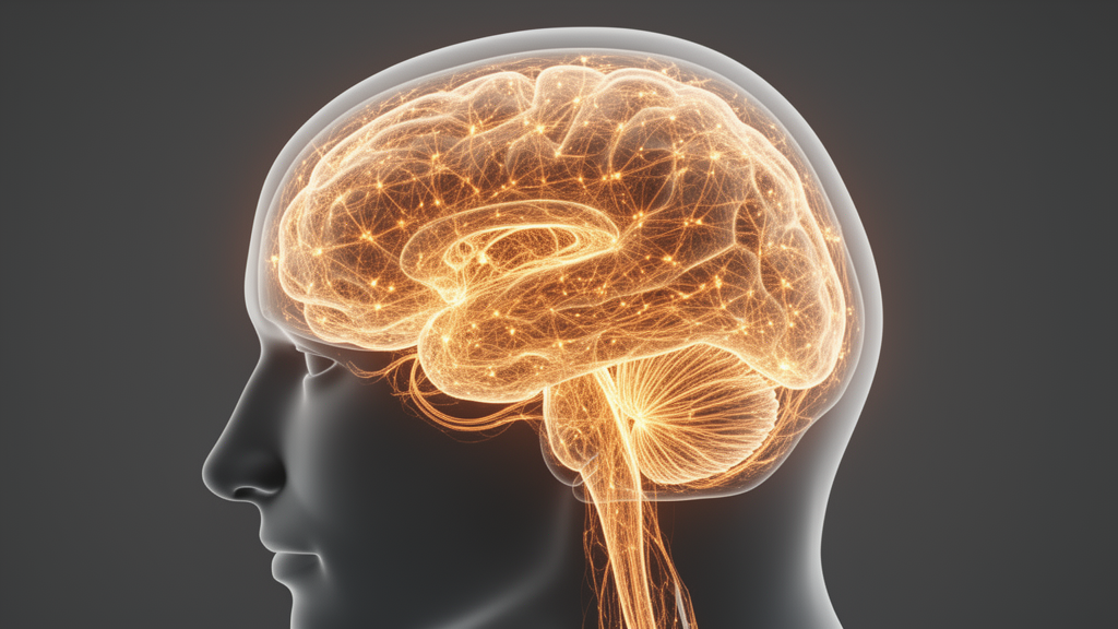
The analysis revealed that electroconvulsive therapy induced prominent changes in specific subcortical and cortical territories. Primary regions of alteration included the bilateral hippocampus and bilateral amygdala, showing consistent shifts across the evaluated psychiatric populations.
Significant changes were also concentrated in right-hemispheric structures, including the right parahippocampal gyrus, the right superior temporal pole, the right anterior cingulate cortex, and the right insula. Additional right-sided changes were identified in the basal ganglia, specifically involving the right lateral globus pallidus, the right medial globus pallidus, and the right caudate body.
Multimodal convergence was particularly prominent within the right hippocampus, underscoring its role as a consistent site of treatment-related neurobiological change. The study authors noted that these convergent structural and functional modifications may relate to therapeutic efficacy as well as adverse cognitive effects observed clinically following electroconvulsive therapy.
By identifying these spatially convergent changes, the novel multimodal analysis advances scientific understanding of the neurobiological substrates altered by electroconvulsive therapy.
Furthermore, the authors emphasized that this work directly informs future investigations into the underlying mechanisms of electroconvulsive therapy.
Subject of Research: Brain structural and functional alterations following electroconvulsive therapy in psychiatric disorders
Article Title: Multimodal alterations of brain structure and function after electroconvulsive therapy in psychiatric disorders: a voxel-based meta-analysis
Article References: Yu, J., Xu, Q., Ma, L., Huang, Y., Zhu, W., Liang, Y., Tang, W., Zhu, C., & Jiang, X. (2026). Multimodal alterations of brain structure and function after electroconvulsive therapy in psychiatric disorders: a voxel-based meta-analysis. Translational Psychiatry. https://doi.org/10.1038/s41398-026-04582-6
Image Credits: AI Generated
DOI: 10.1038/s41398-026-04582-6
Keywords: electroconvulsive therapy, voxel-based morphometry, activation likelihood estimation, neuroimaging, hippocampus, amygdala, psychiatric disorders, Translational Psychiatry, brain structure, brain function, voxel-based pathophysiology, insula, anterior cingulate cortex, parahippocampal gyrus, globus pallidus, caudate body
News Source: Glenn Wilkins. (November 21, 2026). Meta-Analysis Identifies Convergent Brain Alterations Induced by Electroconvulsive Therapy. Scienmag.