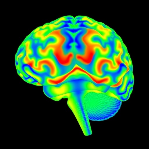
Functional magnetic resonance imaging (fMRI) has ushered in a new era in the field of neuroscience, offering researchers a window into brain activity as it unfolds in real-time. However, while fMRI provides valuable insights into brain function, it primarily reflects changes in blood flow, leaving gaps in our understanding of the underlying neurochemical processes that drive these activations. To bridge this gap, a cutting-edge protocol merging fMRI with electrochemical measurements has been developed, opening doors to a more comprehensive view of brain activity.
This innovative protocol allows for the simultaneous assessment of neurochemical dynamics alongside brain-wide activity patterns. At its core, it integrates fast-scan cyclic voltammetry (FSCV) techniques with fMRI, enabling researchers to measure neurochemical signals—specifically, the dynamics of dopamine in this instance—while observing the broader context of brain function through fMRI. The combination of these two advanced methodologies provides a synergistic approach to understanding the complex interplay of neurotransmitters and neural activity.
One of the primary challenges in this type of multimodal research has been the interference that can arise from synchronization between overlapping datasets. The protocol addresses these challenges head-on, ensuring that the distinct signals captured by each modality do not conflict with or obscure one another. By developing magnetic resonance-compatible electrode designs and optimizing data acquisition settings, researchers can synchronize their measurements with remarkable precision.
To illustrate the practical application of this protocol, the authors showcase in vitro and in vivo procedures for assessing dopamine levels in a flow-cell setup or directly in live rats during MRI scans. Dopamine, a critical neurotransmitter involved in reward pathways, movement, and various neuropsychiatric conditions, serves as an exemplary target to explore under this integrated approach. This demonstration not only highlights the protocol’s feasibility but also its potential in elucidating the neurochemical basis of behaviors and cognitive processes.
The procedural details provided in the protocol are extensive, designed for researchers who possess expertise in MRI, FSCV, and stereotaxic surgeries. This level of detail ensures reproducibility and allows for the protocol to be adapted for other analytes that can be measured using FSCV or related techniques, such as amperometry and aptamer-based sensing. By offering clear, step-by-step guidance, it paves the way for future studies of neurovascular coupling that also consider the intricate neurochemical landscape in which brain networks operate.
The implications of successfully implementing this protocol are profound. Researchers can begin to dissect the intricate relationships between neural activity and neurotransmitter release, potentially leading to breakthroughs in our understanding of brain disorders. For instance, dysregulations in dopamine signaling have been implicated in various conditions, including Parkinson’s disease, schizophrenia, and addiction. By capturing data on both neuron firing and the corresponding neurochemical output, scientists stand to gain insights that could translate into novel therapeutic approaches.
Moreover, this methodology could greatly enhance the study of neurovascular coupling—the relationship between neuronal activity and cerebral blood flow. Historically, this coupling has been challenging to study directly due to the limitations of isolated measurement methods. The protocol integrates different modalities, giving researchers the ability to see brain activity and the neurochemical changes that accompany it, thereby creating a more holistic view of brain function.
Applications of this technology extend far beyond basic neuroscience research. As pharmacological treatments often target specific neurochemical pathways, this technique opens up avenues for translational research. In clinical settings, understanding how drugs interact with neurotransmitter systems in real-time could inform treatment protocols, refine therapeutic strategies, and improve patient outcomes for individuals suffering from psychiatric and neurological disorders.
Additionally, the potential for using this protocol in animal research studies lays the groundwork for future human studies. As researchers build a robust dataset of neurochemical dynamics in animal models, they can lay the foundation for translational research that might apply the insights gained to human patients. The clinical applications of this methodology could eventually contribute to personalized medicine approaches in treating neuropsychiatric conditions by tailoring interventions based on real-time neurochemical feedback.
The timeline for implementing this protocol is also relatively manageable, with the whole process estimated to be completed in a week. This expeditious timeframe is critical in a field that often faces delays due to the complexities associated with integrating multiple technologies. The speed at which researchers can transition from method development to actual experimentation may accelerate discoveries in the field considerably.
The groundwork laid by this protocol marks a significant advancement in neuroscience, where the convergence of technologies can unravel the complexities of brain function. By harmonizing electrochemical measurements with functional imaging, researchers can derive a nuanced understanding of both local and network-level brain processes. Ultimately, the integration of these modalities empowers neuroscientists to piece together the puzzle of brain mechanisms, ultimately advancing our knowledge and treatment of brain disorders.
In conclusion, this novel approach to assessing neurochemical signals through FSCV in conjunction with fMRI is a game-changer for neuroscience research. It opens new avenues for exploring the interactions between neurotransmitters and brain activity, thus providing a richer, multidimensional perspective on brain function. Future studies leveraging this protocol could unveil transformative insights regarding neurophysiology, potentially leading to innovative solutions for clinical challenges in neuroscience.
Subject of Research: Integration of electrochemical measurements with functional MRI.
Article Title: Measurement of electrochemical brain activity with fast-scan cyclic voltammetry during functional magnetic resonance imaging.
Article References:
Shnitko, T.A., Walton, L.R., Peng, TY.R. et al. Measurement of electrochemical brain activity with fast-scan cyclic voltammetry during functional magnetic resonance imaging.
Nat Protoc (2025). https://doi.org/10.1038/s41596-025-01250-9
Image Credits: AI Generated
DOI:
Keywords: neurochemistry, brain imaging, functional MRI, fast-scan cyclic voltammetry, neurotransmitters, neuroscience research.
Tags: advanced brain imaging techniquesbridging gaps in brain activity understandingchallenges in synchronizing neuroimaging datacomprehensive brain activity mappingelectrochemical measurements in fMRI studiesfMRI and fast-scan voltammetry integrationinnovative protocols in neuroimagingmultimodal neuroscience research methodsneurochemical dynamics in brain activityreal-time brain function assessmentsynergistic approaches in neuroscienceunderstanding dopamine signaling in the brain


