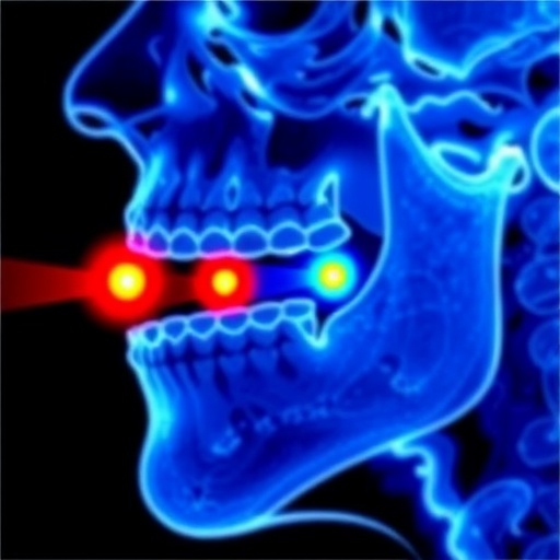
A groundbreaking multicenter clinical trial is poised to revolutionize the staging and treatment of early-stage oral squamous cell carcinoma (OSCC) by introducing a novel magnetic sentinel lymph node (SLN) detection method that leverages superparamagnetic iron oxide nanoparticles (SPIO). This advanced approach addresses critical limitations inherent in the traditional use of radioactive tracers, ushering in a new era of precision oncology and diagnostic accuracy.
The conventional technique for SLN biopsy in OSCC typically relies on tracers labeled with technetium-99m (99mTc), a radioactive isotope. Despite its widespread use, this method suffers from a phenomenon known as “shine-through,” where the intense uptake of radioactive tracer at the primary tumor injection site creates a radiographic glow that obscures sentinel lymph nodes situated in close proximity. This challenge is especially pronounced in floor of mouth tumors given the anatomical closeness of the nodes, often leading to difficulties in accurate lymphatic mapping and increasing the risk of false-negative findings.
Researchers behind the multicenter MAGNETICS trial have devised a comprehensive magnetic SLN biopsy technique combining peritumoral injection of SPIO with magnetic resonance imaging (MRI)-enhanced lymphography and intraoperative magnetometer-guided node detection. SPIO nanoparticles, by virtue of their superparamagnetic properties, provide a distinct imaging signature. This not only circumvents the shine-through artifact associated with radioisotopes but also offers superior anatomical detail by highlighting lymphatic drainage pathways with high spatial resolution on MR lymphography scans, paving the way for more precise surgical navigation.
The trial’s methodology involves enrolling 82 patients diagnosed with early-stage OSCC who will undergo transoral tumor resection alongside dual-modality SLN biopsy. Patients will receive the SPIO injection in addition to the conventional radioactive [99mTc]Tc-nanocolloid tracer combined with indocyanine green dye. This dual labeling strategy enables direct head-to-head comparison of sensitivity, negative predictive value, and interobserver reliability between the magnetic and standard approaches, providing robust clinical evidence regarding the diagnostic performance of the magnetic technique.
One of the pivotal advantages of SPIO-enhanced SLN mapping lies in eliminating patients’ exposure to ionizing radiation. Given growing concerns about cumulative radiation doses from nuclear medicine procedures, this innovation heralds a safer diagnostic process, potentially reducing long-term carcinogenic risks and improving patient comfort and compliance. Moreover, MRI’s unparalleled soft tissue contrast facilitates accurate anatomical localization of sentinel nodes relative to critical neurovascular structures, enhancing surgical precision and preserving function.
The intraoperative detection of SLNs employing a handheld magnetometer further enhances the surgeon’s ability to pinpoint sentinel nodes labeled with SPIO particles. This magnetic guidance offers real-time feedback without reliance on gamma counters or fluorescence detection systems, simplifying the operative workflow and potentially lowering costs and logistical burdens associated with radiotracer handling and waste disposal.
Preliminary evidence from pilot studies suggests that magnetic SLN biopsy may reduce false-negative rates, a significant concern where missed metastatic nodes can lead to understaging and affect prognosis adversely. By capturing sentinel nodes obscured due to shine-through or anatomical variants, the magnetic method promises to refine the accuracy of nodal staging in OSCC, which directly impacts therapeutic decision-making and patient outcomes.
Beyond OSCC, this magnetic SLN detection approach holds promise for expanding into other malignancies where sentinel node assessment is critical, including breast cancer and melanoma. Its radiation-free profile and enhanced detection capabilities could reshape standard practices across oncologic surgery, heralding broader applications in personalized cancer care.
The trial’s multicenter design bolsters the generalizability of findings across various clinical settings and patient demographics, ensuring that results reflect practical utility and scalability. Rigorous assessment includes evaluating patient perspectives, acknowledging that innovations in diagnostic techniques must align with patient comfort and preferences to achieve widespread adoption.
Ethical oversight and regulatory approvals underpin the trial’s conduct, with authorization granted by the Medical Research Ethical Committee NedMec (number: 2023/157) and registration in the Netherlands Trial Register (NL81165.041.22), aligning the study with international clinical research standards and transparency mandates.
If proven successful, the magnetic SLN biopsy procedure will establish a new standard for lymph node staging in OSCC, mitigating current limitations while enhancing diagnostic confidence. This, in turn, could lead to more tailored surgical interventions, minimizing overtreatment and preserving quality of life without compromising oncologic safety.
The magnetic approach’s integration with advanced MRI lymphography also exemplifies the growing convergence of nanotechnology, imaging innovations, and surgical oncology, illustrating how interdisciplinary collaborations can drive transformative advances in cancer diagnostics and therapeutics.
Moreover, the trial highlights the importance of imaging biomarkers in guiding precision surgery, emphasizing that future cancer care will increasingly depend on sophisticated, non-invasive detection tools capable of mapping disease spread with unparalleled accuracy.
As clinicians await the results of this pivotal study, the potential for magnetic sentinel lymph node detection to replace radioactive tracers invites a paradigm shift in how early-stage OSCC is managed worldwide, offering a radiation-free, highly sensitive, and patient-friendly alternative that may ultimately improve survival and reduce morbidity.
This ambitious endeavor marks a significant milestone in the quest to harness nanotechnology’s diagnostic potential, setting the stage for ongoing innovations that transcend oral cancer and resonate across fields that depend on sentinel node biopsy for staging and treatment planning.
In summary, the multicenter MAGNETICS trial represents a visionary leap towards optimizing cancer diagnostics through magnetic technologies, potentially transforming surgical oncology protocols and enhancing patient outcomes in early-stage oral squamous cell carcinoma and beyond.
Subject of Research: Early-stage oral squamous cell carcinoma sentinel lymph node detection using superparamagnetic iron oxide nanoparticles
Article Title: Magnetic sentinel lymph node detection using superparamagnetic iron oxide in early-stage oral squamous cell carcinoma: design and rationale of the multicenter magnetics trial – study protocol
Article References:
Donders, D.N.V., Heldens, G.T.N., Tellman, R.S. et al. Magnetic sentinel lymph node detection using superparamagnetic iron oxide in early-stage oral squamous cell carcinoma: design and rationale of the multicenter magnetics trial – study protocol. BMC Cancer 25, 1539 (2025). https://doi.org/10.1186/s12885-025-14866-7
Image Credits: Scienmag.com
DOI: https://doi.org/10.1186/s12885-025-14866-7
Tags: diagnostic accuracy in cancer treatmentearly-stage cancer staging techniquesintraoperative magnetometer-guided detectionlymph node biopsy innovationsmagnetic sentinel node detectionMRI-enhanced lymphographymulticenter clinical trial resultsnovel cancer detection methodsoral squamous cell carcinoma treatmentprecision oncology advancementsshine-through phenomenon in imagingsuperparamagnetic iron oxide nanoparticles

