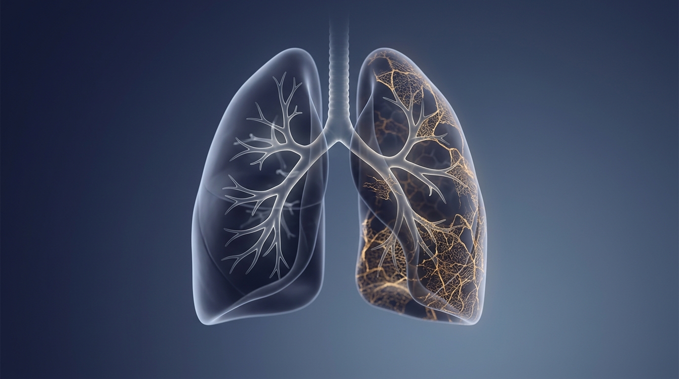
More than one in three survivors of COVID-19-related acute respiratory distress syndrome left the intensive care unit with fibrotic changes visible on chest CT six months after discharge, according to a large French and Belgian study published in Annals of Intensive Care. Among 440 patients analysed from the RECOVIDS cohort, 162 patients, or 36.8 percent, showed pulmonary fibrotic changes at follow-up, defined as interstitial lesions with volume loss, architectural distortion, traction bronchiectasis or honeycombing. The findings come from a prospective observational substudy conducted across 32 centres, 30 in France and 2 in Belgium, and represent what the authors describe as the largest cohort of COVID-19 ARDS survivors evaluated by chest CT at a median interval of 6.5 months after ICU discharge.
The study enrolled adult patients admitted to the ICU with SARS-CoV-2 infection confirmed by polymerase chain reaction between September 2020 and June 2021. To qualify, patients had to meet the Berlin 2012 definition of ARDS or have received high-flow nasal oxygen with a flow of at least 50 litres per minute, an inspired oxygen fraction above 50 percent and a PaO2/FiO2 ratio of 200 or less. Of the 555 patients included in the parent RECOVIDS study, 94 were excluded for substudy-specific reasons such as failure to attend follow-up CT or a history of interstitial lung disease, and a further 21 were excluded as protocol violations, leaving 440 patients for analysis.
Follow-up imaging was scheduled at 6 plus or minus 1 months after ICU discharge. Two thoracic radiologists independently assessed baseline and follow-up scans, resolving disagreements in a joint third review session. The team applied fibrotic terminology from the Fleischner Society glossary to describe baseline features, while acknowledging that descriptors such as traction bronchiectasis may be reversible in the acute setting and not indicative of established fibrosis. Seemingly pre-existing subpleural interstitial abnormalities were not counted as fibrotic changes unless progression was confirmed by comparing both scans. Lesion extent was quantified both visually, as an average percentage score from 0 to 100, and automatically using the CT Pneumonia Analysis application on Syngo.Via software.
By multivariable analysis, the odds of fibrotic changes at follow-up were significantly higher in older patients, those with a body mass index below 30 kilograms per square metre, those with a Charlson comorbidity score of one or higher at ICU admission, those who required invasive mechanical ventilation during the ICU stay, and those showing at least two early fibrotic signs and greater lung involvement on the baseline CT. Each additional year of age and each additional percent of lung involvement on the initial scan raised the odds of developing fibrotic changes at six months by roughly 4 percent and 3 percent respectively. Early fibrotic signs on the baseline scan carried an adjusted odds ratio of 3.7, with a 95 percent confidence interval of 1.5 to 10, for predicting later fibrotic changes.
Patients who went on to develop fibrotic changes were significantly older, more often men, less often obese, more frequently had a Charlson score of one or higher and had higher median Simplified Acute Physiology Score II values than patients without fibrosis. They also more often showed early fibrotic signs at baseline, 46 percent versus 29 percent, and had more extensive lung involvement, measured visually as a median 51.8 percent of lung affected compared with 37.5 percent in the non-fibrotic group, or 57.3 percent versus 44.3 percent by automated analysis. Notably, early fibrosis was absent in more than half of the patients who later developed fibrotic changes, while these signs regressed in fewer than a third of those who remained free of fibrosis, underscoring both the potential for repair and the risk of scarring emerging later during the ICU stay.
To translate these predictors into practice, the researchers built a nomogram combining age, sex, body mass index, Charlson comorbidity score, invasive ventilation status, early fibrotic signs and baseline lung involvement. The tool yielded an area under the curve of 80.6 percent, with a 95 percent confidence interval of 76.4 to 84.8, for the entire cohort, and internal validation by cross-validation gave areas under the curve of 81.2 for the training set and 79.8 for the test set. A worked example in the paper illustrates its use: a 60-year-old man with a body mass index below 30, a Charlson score of 1, invasive mechanical ventilation, two early fibrotic signs and 55 percent lung involvement on baseline CT would carry an estimated 74 percent risk of fibrotic changes within six months.
The pattern of scarring varied. Late organizing pneumonia, with or without distortion, was the most frequent radiological pattern overall, seen in 206 patients, or 47.2 percent of the cohort, a finding the authors suggest may correspond to the specific residual impact of the virus, since ground glass opacities and consolidations tend to regress by one year. Meanwhile, 30 patients, about 19 percent of the 160 evaluable patients with fibrotic changes, showed mainly anterior fibrosis compatible with post-ventilatory changes. The authors link this anterior distribution to ventilator-induced lung injury: during ARDS, consolidation of the dependent posterior lung regions leaves the anterior zones most exposed to overdistension during invasive ventilation. Consistent with this, the duration of invasive ventilation and the proportion of tracheostomy for weaning were significantly higher in the fibrotic group, and median airway plateau pressure in that group was 30 centimetres of water, a threshold beyond which the risk of ventilator-induced injury is considered significant, although the difference did not reach statistical significance. Prone positioning showed no protective effect against fibrotic changes in this cohort.
The clinical footprint of the scarring was measurable. Patients with fibrotic changes more frequently reported shortness of breath on the modified Medical Research Council dyspnea scale and had significantly lower oxygen saturation at rest and after exertion. Pulmonary function testing revealed a restrictive respiratory profile with significantly impaired forced vital capacity, total lung capacity, diffusion capacity for carbon monoxide and carbon monoxide transfer coefficient. On the six-minute walk test, patients with fibrosis covered 29 metres less on average, a difference that bordered on statistical significance but was judged clinically relevant, particularly as it was not explained by the higher prevalence of emphysema in the fibrotic group. Residual lesions in the fibrotic group affected a median of 14 percent of the lungs, exceeding the 10 percent threshold proposed to deem lesions symptomatically significant. Quality of life, assessed with the modified 36-item Short Form Health Survey, was more impaired in the physical domain among patients with fibrosis, with no differences in the physical or mental summary scores or in the Visual Simplified Respiratory Questionnaire.
One counterintuitive result concerned body weight. Obesity, defined as a body mass index above 30, appeared protective against fibrotic changes despite its association with an increased risk of COVID-19 ARDS. The authors offer a physiological explanation: in obese patients ventilated invasively, hypoxemia often results from atelectasis and ventilation-perfusion mismatch, which may lead clinicians to overestimate ARDS severity in collapsed but unharmed lung areas that leave no scarring once the collapse resolves. Inflammatory and metabolic factors linked to obesity could also account for lesser lung involvement and ultimately fewer sequelae. The protective effect of obesity on mortality in non-COVID-19 ARDS, previously termed the obesity paradox, remains uncertain in COVID-19 ARDS, since one meta-analysis found no significant survival impact.
The authors caution that their model may not generalise to ARDS of other causes because of the specificities of COVID-19 ARDS, and that fibrotic changes on CT cannot be ruled out as having preceded the infection, although exclusion of patients with advanced respiratory histories and seemingly pre-existing interstitial abnormalities limited this possibility. Evaluation at six months may also overestimate fibrosis, since some residual non-fibrotic lesions may still improve over time, although current data suggest fibrotic changes remain stable, and studies have confirmed that changes identified at six months persisted up to 24 months after COVID-19 ARDS. It is also difficult to separate symptoms arising from fibrosis from those of post-intensive care syndrome, since patients with fibrosis more often required invasive ventilation and had hospital stays more than twice as long. The authors suggest that scans performed more than five days before ICU admission may underestimate lesion burden, with maximal quantification occurring between five and ten days, and argue that early identification of high-risk patients could enable tailored follow-up and, potentially, antifibrotic treatment.
Subject of Research: Predictive factors for pulmonary fibrotic changes six months after ICU discharge in survivors of COVID-19-related acute respiratory distress syndrome
Article Title: ICU predictive factors of fibrotic changes following COVID-19 related ARDS: a RECOVIDS substudy
Article References: Demeyere, M., Fournel, I., Sow, A.-K., Gélinotte, S., Nyunga, M., Berraies, A., Labruyère, M., Ampere, A., Sauneuf, B., Daubin, C., Delbove, A., Badie, J., Bulpa, P., Delacour, D., Lefevre, C., Nseir, S., Artaud-Macari, E., Ramakers, M., Bironneau, V., … Declercq, P.-L. (2025). ICU predictive factors of fibrotic changes following COVID-19 related ARDS: a RECOVIDS substudy. Annals of Intensive Care, 15(1), Article 177. https://doi.org/10.1186/s13613-025-01577-2
Image Credits: AI Generated
DOI: 10.1186/s13613-025-01577-2
Keywords: COVID-19, ARDS, pulmonary fibrosis, intensive care, chest CT, nomogram, invasive mechanical ventilation, organizing pneumonia, pulmonary function tests, RECOVIDS study, ventilator-induced lung injury, obesity paradox
News Source: Ophelia Keating. (November 4, 2026). Lung scarring strikes a third of COVID-19 ARDS survivors six months after ICU discharge. Scienmag.