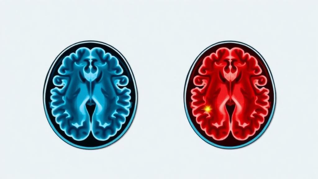
A team of researchers from Daffodil International University in Bangladesh, working with colleagues at Multimedia University in Malaysia, has unveiled a compact artificial intelligence system that classifies brain tumors on MRI scans with 99.2 percent accuracy while telling clinicians how confident it is in every single prediction. The framework, called the Hybrid Scale-Attention Network, was described in the journal Discover Artificial Intelligence and is notable not only for its near-perfect benchmark scores but for the unusual honesty of its engineering: the authors openly report where their model falls short of rivals, which design ideas they tested and discarded, and why statistical significance is hard to claim when a benchmark is nearly saturated.
The clinical motivation is stark. The 2021 World Health Organization classification describes more than 100 distinct tumor entities of the central nervous system, and even with intensive multimodal treatment the five-year survival rate for high-grade gliomas remains below 10 percent. Interpreting MRI scans is time-consuming, and inter-observer variability among radiologists can reach 30 percent in challenging cases, while specialist expertise is unevenly distributed across the globe. Deep learning has repeatedly posted impressive numbers on standardized brain tumor benchmarks, yet three persistent gaps have blocked clinical translation: models are computationally heavy, they are rarely validated on data from different scanners and protocols, and their softmax outputs are notoriously overconfident on ambiguous or out-of-distribution inputs.
The new architecture attacks all three weaknesses at once. Its backbone is EfficientNet-B3, pretrained with the Noisy Student semi-supervised procedure on ImageNet-1K and the unlabeled JFT-300M corpus, which contributes roughly 10.28 million of the model’s 10.97 million total parameters. That figure is 7.8 times smaller than the 86 million parameters of ViT-B/16 and about 92 percent smaller than VGG-16’s 134 million, making the system plausible for deployment in resource-constrained clinical settings where giant models are impractical.
The architectural centerpiece is the Adaptive Multi-Scale Module, or AMSM, inserted after each of three hierarchical feature stages of the backbone. Tumors vary enormously in spatial extent: an infiltrative glioma spreads across a wide region, while a small pituitary adenoma may occupy only a few pixels in the sellar region. AMSM handles this diversity with three parallel depthwise-separable dilated convolutional branches at dilation rates 1, 2, and 4, each enlarging the receptive field without losing resolution, plus an identity branch that preserves the unmodified features. A small two-layer network reads globally pooled descriptors of all four branches and emits softmax gating weights that sum to one, so the fused output is a learned convex combination of multi-scale evidence rather than a fixed aggregation. Because depthwise-separable factorization requires only C times the sum of the kernel size squared and the channel count, instead of the channel count squared times kernel size squared of a standard convolution, the module costs roughly 6.6 times fewer parameters than equivalent conventional branches: 692,490 parameters instead of 4,546,320.
The second innovation concerns trust. Instead of a plain softmax head, the network ends in an Evidential Deep Learning classifier that recasts classification as estimation of a Dirichlet distribution. Logits pass through a softplus function to produce non-negative evidence, Dirichlet parameters are set to evidence plus one, and the uncertainty mass, or vacuity, is defined as the number of classes divided by the Dirichlet strength. Large accumulated evidence yields low uncertainty; thin evidence yields a flag. Training combines an evidential cross-entropy built on the digamma function, a KL divergence term that pulls non-evidence entries toward a uniform Dirichlet prior and is annealed over the first ten epochs, and a focal correction with class weights to handle imbalance. The result is deterministic, single-pass uncertainty quantification at an inference cost of 16.8 milliseconds per image, roughly 20 times faster than MC-Dropout with 20 stochastic forward passes, which took 340.5 milliseconds.
Performance on the primary benchmark, BRISC 2025, a 6,000-image expert-annotated T1-weighted dataset derived from BraTS 2021 with official splits of 5,000 training and 1,000 test images, was 99.20 percent accuracy and 99.30 percent macro-F1, with an AUC-ROC of 0.9995 and only 0.95 gigaflops of compute. Five-fold cross-validation returned a mean accuracy of 99.06 percent with a standard deviation of 0.17 percent, and bootstrap confidence intervals on the test set spanned 98.6 to 99.7 percent. Of the eight errors among 1,000 test images, seven carried uncertainty scores in the 90th percentile of the test distribution, and the model achieved perfect recall on the No Tumor class, meaning no normal scan was falsely flagged as cancerous. Misclassified samples carried 2.6 times higher mean uncertainty than correctly classified ones, and the misclassification detection AUROC reached 0.9461.
Perhaps the most scientifically interesting result is a negative one. During development the team built a Dual Attention Module combining efficient channel attention and spatial attention and tested four architectural variants. In isolation the attention module improved in-domain calibration and achieved the highest uncertainty separation ratio at 2.94 times, but stacking it after AMSM degraded both in-domain accuracy and, more damagingly, cross-dataset generalization on the external PMRAM-BD cohort, a brain MRI collection from Bangladeshi clinical centers acquired under a different protocol. The authors concluded that channel-spatial reweighting after scale-gated fusion encourages dataset-specific feature emphasis at the expense of the domain-invariant spatial representations that AMSM captures, and they excluded the module from the deployed model. It is a rare public example of a deep learning paper arguing against adding more attention.
Cross-dataset evaluation is where the work departs from most of the literature. Without any fine-tuning, the model trained only on BRISC 2025 scored 97.41 percent on PMRAM-BD, a generalization gap of 1.79 percentage points that is smaller than those of ResNet-50, VGG-16, and the EfficientNet-B3 backbone alone, though not smaller than those of ViT-B/16, Swin-Tiny, or DenseNet-121, which achieved the highest absolute external accuracy of 98.07 percent in a single run. The authors are careful here: they frame their model as a strong external performer rather than the best external generalizer, and multi-seed testing showed DenseNet-121’s external advantage was fragile, dropping to 94.88 percent when averaged over three random seeds while the proposed model stayed above 96.35 percent. VGG-16 fared worst, losing 5.94 percentage points, evidence that sheer parameter count does not buy cross-domain robustness. Notably, mean predictive uncertainty rose significantly on the external cohort, with a rank-sum test p-value of 0.000064, showing the model signals reduced confidence exactly when it faces an unfamiliar imaging distribution.
The paper is equally candid about limitations. McNemar’s paired tests against six baselines yielded no statistically significant differences after Holm-Bonferroni correction, which the authors attribute to the near-ceiling performance: at 98.7 to 99.2 percent accuracy on a balanced 1,000-sample test set, only 6 to 11 prediction pairs differ between any two models, so the test cannot resolve gaps below roughly half a percentage point. The evidential model’s expected calibration error of 0.0263 is higher than the softmax baselines, a known property of Dirichlet-parameterized models under standard confidence binning. Classification is performed on 2D slices without volume information, both datasets lack patient-level identifiers so all metrics are image-level, and inference at 19.2 milliseconds per image is slower than ResNet-50 because of an inefficient CUDA implementation of depthwise convolution rather than higher computational load. Interpretability comes from UA-GradCAM heatmaps and a novel Multi-Scale Attention Decomposition that logs the gating weights per class: glioma predictions preferentially activate the rate-4 dilated branch, consistent with infiltrative growth, while pituitary predictions favor the rate-1 branch, consistent with small focal lesions, a pattern the authors compare to a radiologist reporting whether local enhancement or regional mass effect supports a diagnosis.
The authors position the system for retrospective clinical investigation rather than immediate deployment, noting that prospective validation across multiple sites with radiologist consensus as ground truth remains necessary. Even so, the combination of near-state-of-the-art accuracy, a 10.97 million parameter footprint, deterministic single-pass uncertainty, and rigorous multi-cohort evaluation offers a template for what clinically credible medical AI should look like. The code, notebooks, and experimental results are publicly available on GitHub, and the open datasets, BRISC 2025 on Kaggle under CC BY 4.0 and PMRAM-BD on Mendeley Data, mean any laboratory can reproduce the numbers. In a field often criticized for leaderboard chasing and overconfident claims, a model that knows what it does not know, and a paper that says so, may be the most viral result of all.
Subject of Research: Uncertainty-aware deep learning classification of brain tumors from MRI scans
Article Title: A hybrid scale attention network for uncertainty aware brain tumor MRI classification
Article References: A hybrid scale attention network for uncertainty aware brain tumor MRI classification. (n.d.). https://doi.org/10.1007/s44163-026-02386-7
Image Credits: AI Generated
DOI: 10.1007/s44163-026-02386-7
Keywords: brain tumor, MRI, deep learning, uncertainty quantification, evidential deep learning, EfficientNet, multi-scale fusion, attention mechanisms, cross-dataset validation, medical imaging AI, lightweight neural network, interpretable AI
News Source: Cassandra Pierce. (October 14, 2026). Lightweight AI Reads Brain Tumor Scans and Knows When It Is Unsure. Scienmag.