


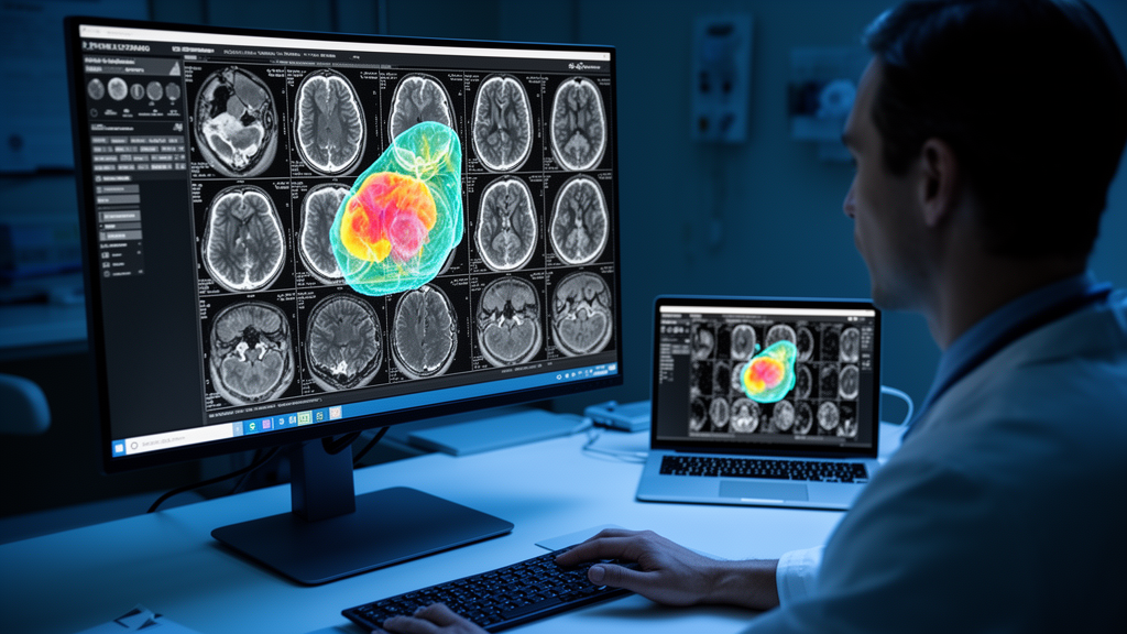
Researchers have unveiled a new artificial intelligence model designed to automatically outline brain tumors and their surrounding swelling on MRI scans, and the numbers behind it are turning heads. The model, called AGDE-Net, short for attention-guided dual-encoder network, was developed to segment two critical regions in adult glioma patients: the tumor core and the peritumoral edema, the fluid-filled tissue that often surrounds a growing tumor. The work, published in the Egyptian Journal of Radiology and Nuclear Medicine, addresses one of the most persistent challenges in medical image analysis: how to combine the strengths of two very different neural network architectures without making the resulting model too heavy for real-world clinical use.
Glioma is the most common malignant brain tumor in adults, accounting for roughly 75 percent of primary brain tumors. Its location, the extent to which it spreads, and the presence of associated edema all have a major impact on patient well-being, producing symptoms that range from headaches and seizures to aphasia and hemiparesis. Treatment decisions, whether maximum safe resection or radiation therapy, depend on precisely delineating where the tumor ends and where the edema begins. That delineation is currently done manually by radiologists working with multimodal MRI, which typically includes T1-weighted, T1-weighted contrast-enhanced, T2-weighted, and T2-weighted FLAIR sequences. Manual segmentation is time-consuming, varies between observers, and is vulnerable to fatigue-induced errors, creating a growing demand for automated tools that can enhance diagnostic efficiency and ensure consistency.
The core technical problem the researchers set out to solve lies in how modern segmentation networks blend two complementary types of information. Convolutional neural networks, or CNNs, excel at extracting local spatial patterns and fine-grained structural details, but their limited receptive fields make it hard to model long-range dependencies across an image. Transformer-based architectures, which power much of today’s artificial intelligence, are far better at capturing global context, yet they can dramatically increase computational complexity and memory consumption. Hybrid CNN-Transformer models have emerged as a compromise, but in many existing designs the interaction between the two branches is handled through direct addition, concatenation, or stage-specific aggregation. Those strategies may not properly account for the different contributions of local and global information at corresponding feature resolutions, and Transformer branches can still impose a heavy computational burden.
AGDE-Net tackles this with a dual-encoder design built on the U-shaped network foundation. A CNN encoder, modeled on the original U-Net, captures local and detailed information through four convolutional blocks with downsampling. In parallel, a hierarchically structured Transformer encoder generates multi-level global features at progressively reduced resolutions, from one-half down to one-sixteenth of the original slice size. The Transformer branch borrows a sequence-reduction strategy from existing efficient Transformer designs, applying stage-wise spatial reduction ratios of 1, 2, 4, and 8 to the key and value representations in self-attention. This cuts the computational complexity of self-attention from quadratic in sequence length down to a reduced form, keeping the architecture lightweight. The two branches then feed into an attention-guided feature fusion module, or AGFF, which adaptively modulates and integrates the complementary representations before they pass to a shared decoder, rather than relying on simple addition or concatenation.
To train and evaluate the model, the team used the publicly available BraTS 2025 pre-treatment adult glioma dataset as their primary source. A total of 118 follow-up scans from patients in the training cohort were excluded from model development and reserved for longitudinal robustness evaluation. The remaining 1133 cases were randomly split at the patient level into training, validation, and internal test sets in a 6:2:2 ratio, with no patient overlap between subsets. The original BraTS annotations were converted into two categories: the non-enhancing and necrotic tumor regions combined with the enhancing tumor formed the tumor core, while the edema region served as the peritumoral edema target. For external validation, the researchers included a cohort of 36 pre-treatment glioma patients from Shandong Cancer Hospital, with scans hand-annotated by two radiologists with 8 and 5 years of tumor diagnosis experience and final review by two senior radiologists with 15 and 35 years of experience.
The results were striking. On the internal testing set, AGDE-Net achieved 3D Dice scores of 0.8382 for the tumor core and 0.8044 for the peritumoral edema. For tumor segmentation, it recorded a positive predictive value of 0.8870, a sensitivity of 0.8380, and a 95% Hausdorff distance of 8.0545 millimeters, ranking highest among compared models for Dice, PPV, and boundary accuracy. For edema, it scored a PPV of 0.8172, a sensitivity of 0.8193, and an HD95 of 9.3125 millimeters. The model was benchmarked against seven established segmentation networks, including Swin U-Net, U-Net++, Dense U-Net, Attention U-Net, standard U-Net, H2Former, and Mamba-Unet, all trained under the same loss function and optimizer settings. Paired Wilcoxon signed-rank tests with Holm correction for multiple comparisons showed that AGDE-Net’s Dice improvements over every baseline were statistically significant at p less than 0.05.
Performance held up under more demanding conditions. On the 118 reserved longitudinal follow-up scans, which capture how tumors change over time within the same patients, AGDE-Net achieved the highest Dice scores of 0.8449 for tumor and 0.8374 for edema, along with a tumor sensitivity of 0.8887. H2Former did obtain a lower mean HD95 for edema, at 9.1584 versus 10.9760 millimeters, indicating better boundary agreement on that specific metric, but AGDE-Net maintained superior overlap and lesion-detection performance overall. On the external dataset from Shandong Cancer Hospital, a genuine test of generalization across different scanners and patient populations, the model recorded Dice scores of 0.7935 for tumor and 0.7798 for edema, only minor reductions from internal testing. Qualitative review did reveal failure cases: the model sometimes missed small, low-contrast tumor regions on T1CE images and occasionally misclassified non-edematous regions as edema on FLAIR sequences.
Computational efficiency may prove to be the model’s most clinically relevant feature. AGDE-Net required only 10.027 million parameters and 5.943 billion floating-point operations, with an inference time of 4.91 milliseconds and a memory footprint of 68.05 megabytes for a single-slice input combining all four MRI sequences, measured on an NVIDIA RTX 3090 Ti. Among all compared methods, it showed the lowest GPU memory usage while maintaining competitive speed and accuracy. Ablation experiments confirmed that both components of the design contribute meaningfully: replacing the attention-guided fusion with simple addition dropped tumor Dice from 0.8382 to 0.8218 and edema Dice from 0.8044 to 0.7962, while HD95 boundary errors worsened for both targets. This indicates the performance gains are not solely attributable to the dual-encoder structure itself but to the adaptive fusion mechanism layered on top of it.
The researchers are careful to frame these results as promising rather than definitive. In preliminary radiologist-assisted contouring on the external set, many AI-generated contours required only limited manual adjustment, suggesting the model may help reduce segmentation workload, but the authors note that further evaluation using a standardized multi-reader timing protocol is required. Several limitations are acknowledged: the model showed greater performance variability in small lesion regions than in larger ones, the influence of different input resolutions and preprocessing configurations was not systematically explored, and the two-target formulation does not distinguish enhancing from non-enhancing or necrotic tumor components, limiting detailed characterization of intratumoral heterogeneity. Broader multicenter evaluation across different scanners and acquisition protocols is still needed. Even so, the combination of stable accuracy across internal, longitudinal, and external cohorts, together with a footprint light enough for standard clinical hardware, positions AGDE-Net as a candidate for integration into real diagnostic workflows, where every millisecond and every megabyte counts.
Subject of Research: A deep learning model for segmenting glioma tumors and edema on MRI
Article Title: AGDE-Net: a lightweight attention-guided dual-encoder network for tumor core and peritumoral edema segmentation in adult glioma
Article References: Zhou, Z., Liu, H., Wang, M., Song, H., Li, J., Wang, Z., & Lv, H. (2026). AGDE-Net: a lightweight attention-guided dual-encoder network for tumor core and peritumoral edema segmentation in adult glioma. Egyptian Journal of Radiology and Nuclear Medicine, 57(1), Article 214. https://doi.org/10.1186/s43055-026-01880-3
Image Credits: AI Generated
DOI: 10.1186/s43055-026-01880-3
Keywords: glioma, brain tumor segmentation, deep learning, MRI, Transformer, CNN, AGDE-Net, BraTS 2025, medical imaging, computational efficiency, tumor edema, radiology
News Source: Ophelia Keating. (October 30, 2026). Lightweight AI Network Segments Glioma Tumors and Edema on MRI Scans. Scienmag.



