Intense pulsed light, a technology long used by dermatologists to treat rosacea and pigmented lesions, is emerging as a multi-target therapy for stubborn ocular surface diseases, according to a narrative review published on 30 September 2026 in the journal Eye and Vision. The review, authored by Jiayi Yan, Wei Chen, and Jinyang Li, argues that conditions such as refractory meibomian gland dysfunction, Demodex-associated blepharitis, ocular rosacea, recurrent chalazion, and allergic conjunctivitis share convergent pathological features, including chronic inflammation, abnormal lid-margin vascularity, and microbial or parasitic imbalance, and that intense pulsed light can act on several of these drivers at once.
The technology’s route into ophthalmology began with an accidental observation. In 2002, Dr. Toyos reported that patients undergoing facial intense pulsed light treatment for rosacea experienced improvement in their concomitant refractory dry eye. Because the eyelid is an anatomical extension of the facial skin, sharing pathological targets such as abnormal vascular proliferation, chronic inflammation, and microbial imbalance, clinicians began repurposing a device originally designed to occlude abnormal vasculature and remove pigmented targets. Over the following two decades, protocols were gradually optimized for evaporative dry eye, and indications extended to blepharitis and recurrent chalazia.
The review frames the therapy around the principle of selective photothermolysis, in which light of specific wavelengths is preferentially absorbed by chromophores in target tissues and converted into heat. In meibomian gland dysfunction, this photothermal effect reduces the viscosity of inspissated meibum and improves its fluidity within the gland ducts. When followed immediately by mechanical meibomian gland expression, the treatment works through what the authors describe as a thermal liquefaction and mechanical drainage effect. Clinical studies cited in the review support this synergy, reporting a mean improvement of +13.69 in the meibomian gland yielding secretion score at one month post-treatment, although the response varies with disease severity, glandular structural damage, device parameters, and whether expression was performed.
A second proposed mechanism is vascular and anti-inflammatory. Intense pulsed light is absorbed by hemoglobin within pathologically dilated periorbital capillaries, generating heat that induces microthrombi and vascular occlusion, which may reduce the delivery of inflammatory mediators to the ocular surface. At the molecular level, studies have reported marked decreases in interleukin-6, tumour necrosis factor-alpha, C-X-C motif ligand 1, and interferon-gamma, all with P values below 0.001, alongside a reduction in matrix metalloproteinase-9 positivity from 97% to 69% and a concomitant increase in tissue inhibitor of metalloproteinases-1. These cytokine changes correlate with clinical improvements: tear film breakup time prolongation is associated with decreased tumour necrosis factor-alpha, with a correlation coefficient of -0.814, and improvements in gland secretion scores and symptom indices correlate with reduced levels of multiple pro-inflammatory cytokines.
The review also describes antimicrobial and antiparasitic effects. Chromophores in Demodex mite exoskeletons and internal pigments may absorb the light, causing localized temperature increases and thermal injury. In vitro, standard treatment parameters kill live Demodex mites, and clinical studies have reported significant post-treatment decreases in mite counts on the eyelid margin. The light also appears to reduce bacterial load at the eyelid margin by disrupting bacterial cell walls. Because Demodex mites often harbor bacteria and release inflammatory substances including lipases, reducing mite and bacterial burden may decrease local inflammatory stimuli and complement conventional antimicrobial therapies.
A fourth, more speculative pathway involves photobiomodulation. The red and near-infrared portions of the light spectrum penetrate tissue and are absorbed by cytochrome c oxidase in the mitochondrial membrane, potentially improving electron transport chain efficiency and stimulating adenosine triphosphate synthesis. Cellular-level findings using flavoprotein fluorescence technology showed a significant decrease in flavoprotein fluorescence after treatment, from 13,800 plus or minus 3,200 to 10,200 plus or minus 2,800 arbitrary units, consistent with increased mitochondrial metabolic activity and reduced oxidative stress. Microstructural parameters also improved, with meibomian gland acinar unit density increasing from 78.5 plus or minus 15.2 to 96.3 plus or minus 18.7 glands per square millimeter. The authors stress, however, that direct evidence of neuroimmune restoration, neural repair, or tissue regeneration remains limited, and these effects should be interpreted as emerging hypotheses rather than established outcomes.
The strength of evidence differs substantially across disease categories, the review emphasizes. Meibomian gland dysfunction-related evaporative dry eye has the strongest evidence base, supported by randomized controlled trials and meta-analyses, with studies reporting improvements in tear film stability, meibomian secretion quality, lipid layer thickness, inflammatory cytokine profiles, and patient-reported symptoms, especially when combined with gland expression. For Demodex-associated blepharitis, published studies suggest the treatment may reduce mite burden, lid-margin inflammation, hyperemia, and tear film instability, particularly when combined with topical ivermectin, but the evidence remains less robust and further controlled studies are needed. For ocular rosacea, existing studies suggest improvements in tear film stability, symptom scores, and lid-margin vascular signs, yet the evidence is still more limited than for dry eye.
For recurrent or meibomian gland dysfunction-associated chalazion, the treatment has been explored as a non-invasive adjunctive option, with existing studies suggesting it may promote lesion resolution and reduce recurrence, possibly by improving gland function and the eyelid microenvironment. However, most available studies are limited by small sample sizes, retrospective designs, or short follow-up periods, and larger controlled studies are needed before the therapy can be recommended as a standard alternative to established treatments. Allergic conjunctivitis is discussed separately as an emerging adjunctive application: a recent randomized controlled trial suggested that the treatment combined with conventional anti-allergic therapy may improve symptoms and signs of refractory seasonal allergic conjunctivitis in children, but the evidence remains preliminary and its use should currently be considered investigational.
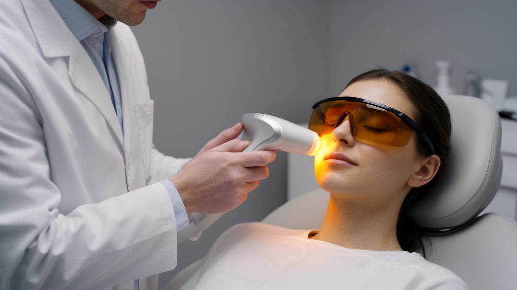
Practical protocol details matter considerably, according to the review. Devices emit polychromatic light in the range of 515 to 1200 nanometers, with filters selecting the desired wavelength; most studies chose 590-nanometer or 560-nanometer filters to treat eyelid-margin telangiectasia. Energy density is typically set according to Fitzpatrick skin phototype, with 12 to 16 joules per square centimeter common for lighter skin types I to III and 9 to 11 joules per square centimeter for darker types IV to V to avoid epidermal damage. A tripulse mode is typically applied, and with complete corneal and scleral coverage using a Jaeger opaque metal eye shield, direct irradiation of the upper eyelid skin at 10 joules per square centimeter has been reported as safe and effective. Standard treatment involves three to four sessions spaced two to four weeks apart, with more sessions, such as eight treatments at three-week intervals, potentially offering longer-lasting improvements in severe or refractory cases. Baseline gland morphology predicts response: patients with severe glandular structural damage, such as high gland loss scores and sequential extreme thinning, respond poorly even when eyelid-margin signs are mild.
The authors are candid about the limitations of the current evidence. Studies differ substantially in device, filter, fluence, pulse mode, treatment interval, number of sessions, adjunctive therapy, outcome measures, and follow-up duration, which limits direct comparisons and complicates the definition of standardized protocols. Follow-up is often limited to weeks or months, so the durability of benefits and the need for maintenance therapy remain unclear, and long-term safety data and high-quality randomized controlled trials remain limited, particularly beyond meibomian gland dysfunction. Practical factors, including cost, device availability, operator experience, and repeated in-office visits, may also limit real-world implementation. The review concludes that intense pulsed light is best understood as a phenotype-directed, mechanism-oriented adjunctive therapy for selected refractory patients rather than a universal treatment for all ocular surface diseases, and that future research should standardize treatment parameters and adverse-event reporting, clarify cellular and molecular mechanisms, and pursue combined energy strategies, phenotype-stratified protocols, and biomarker-driven personalization.
Subject of Research: Intense pulsed light therapy for refractory ocular surface diseases
Article Title: Intense pulsed light as a multi-target intervention paradigm for refractory ocular surface diseases: a review
Article References: Yan, J., Chen, W., & Li, J. (2026). Intense pulsed light as a multi-target intervention paradigm for refractory ocular surface diseases: a review. Eye and Vision, 13(1), Article 41. https://doi.org/10.1186/s40662-026-00510-3
Image Credits: AI Generated
DOI: 10.1186/s40662-026-00510-3
Keywords: intense pulsed light, ocular surface disease, meibomian gland dysfunction, dry eye, Demodex blepharitis, ocular rosacea, chalazion, photobiomodulation, tear film, meibomian gland expression, ophthalmology, review
News Source: Ophelia Keating. (October 28, 2026). Light Therapy Shows Promise for Stubborn Eye Surface Diseases, Review Finds. Scienmag.