The study, published in the journal Discover Sensors, is a theoretical and computational exercise: no physical device was built and no real urine was measured. Even so, the numbers that emerge from the simulations are striking enough to have drawn attention, with the proposed structure reaching a calculated sensitivity of 416.42 degrees per refractive index unit, a figure the authors say outperforms earlier designs by a clear margin.
The sensor follows the Kretschmann configuration, the classic arrangement for surface plasmon resonance sensing, in which p-polarized light strikes a prism and couples into collective oscillations of free electrons at a metal surface. When the angle of incidence matches the momentum of these surface plasmons, reflectance drops sharply, and the position of that dip shifts whenever the refractive index of the liquid touching the sensor changes. Because dissolved glucose raises the refractive index of urine, tracking the resonance angle offers a label-free route to estimating sugar content without reagents, enzymes, or fluorescent tags. The authors modeled the optical response with the transfer matrix method at a wavelength of 633 nanometers, sweeping the incidence angle to locate the resonance minimum for each analyte.
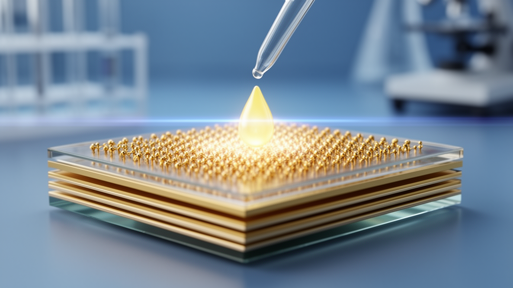
Every layer in the stack was chosen for a specific optical role. Light enters through a K108 (LZOS) prism with a refractive index of 1.5115, which the authors selected to improve coupling efficiency. Beneath it sits a 56-nanometer film of silver, the plasmonic engine of the device, described by a complex refractive index of 0.056206 plus 4.27761i at the operating wavelength. Silver sustains stronger plasmonic oscillations than most metals, but it tarnishes readily, so the design adds dielectric and semiconducting capping layers that both shield the metal from oxidation and sharpen the evanescent field at the sensing interface.
Those capping layers are where the design departs from conventional biosensors. An 8-nanometer titanium dioxide layer, with a refractive index of 2.5836 at 633 nanometers, enhances light trapping near the metal. A 1-nanometer zinc telluride layer, refractive index 2.9887, further tightens the confinement of the optical field. On top sits the active sensing layer: a single 0.53-nanometer monolayer of aluminium antimonide, an III–V semiconductor with a narrow direct bandgap of 1.6 electronvolts and a high refractive index of 3.3. The authors argue that this combination of high refractive index, strong light–matter interaction, and surface activity makes AlSb an effective transducer for biomolecules at the sensing surface.
The optimization was carried out in stages. First, the silver thickness was varied from 40 to 56 nanometers in 2-nanometer steps while the dielectric layers were held at 1 nanometer each; thicker silver produced deeper, narrower resonance dips, and 56 nanometers gave the best resonance characteristics, with sensitivity rising to 144.14 degrees per refractive index unit at that point. Next, the titanium dioxide thickness was swept from 1 to 8 nanometers at fixed silver and zinc telluride thicknesses. Sensitivity climbed steeply with the dielectric layer, from 144.14 to 416.42 degrees per refractive index unit, as plasmonic coupling strengthened. Although the minimum reflectance and the resonance linewidth both increased slightly, the structure still achieved a quality factor of 135.80 per refractive index unit and a figure of merit of 129.07 at the 8-nanometer thickness, which the authors adopted for the final design.
The sensing target was the refractive index window of urine at different glucose loads. The simulations covered analyte refractive indices of 1.335, 1.336, 1.337, 1.338, 1.341, and 1.347, spanning what the authors describe as non-diabetic through highly diabetic urine. Across this range the resonance angle shifted by up to 4.997 degrees for the full five-layer structure, labeled S5, which pairs the K108 prism with silver, titanium dioxide, zinc telluride, and AlSb. The quality factor rose from 102.66 to 142.29 per refractive index unit and the signal-to-noise ratio from 0.10 to 1.71 as the analyte index increased, while the peak figure of merit and combined sensitivity factor reached 98.12 and 95.47 at a refractive index of 1.338.
To test whether AlSb was genuinely the best choice for the top layer, the team benchmarked three alternative two-dimensional materials in the same stack: black phosphorus, graphene, and tungsten disulfide. Black phosphorus reached a sensitivity of 395.07 degrees per refractive index unit with a quality factor of 118.50, graphene managed 320.3 degrees per refractive index unit, and the tungsten disulfide variant delivered 384.4 degrees per refractive index unit over part of the range. AlSb came out ahead on every headline metric, which the authors attribute to its high refractive index and strong plasmonic field enhancement. They also compared the full design against previously published urine-glucose SPR sensors, including gold-graphene, MXene-zinc oxide, black phosphorus, zinc selenide, and aluminum nitride configurations that had reported sensitivities between 133.5 and 394.44 degrees per refractive index unit; the proposed structure exceeded all of them in both sensitivity and quality factor.
Supporting simulations reinforce the physical picture. Finite-difference time-domain calculations of the electric field distribution show strong localization of the electromagnetic field at the silver–dielectric interface, with the titanium dioxide, zinc telluride, and AlSb layers confining the evanescent field close to the sensing surface where analyte molecules interact with it. The field decays exponentially into the sample, as expected for surface plasmon resonance, with a calculated penetration depth of 344.83 nanometers, deep enough for glucose molecules in the adjacent fluid to influence the resonance condition.
The authors also sketch a plausible fabrication route, though it remains untested. The prism surface would be cleaned with methanol, acetone, and deionized water; the 56-nanometer silver film deposited by electron-beam evaporation in high vacuum; the 8-nanometer titanium dioxide added by atomic layer deposition; the 1-nanometer zinc telluride applied by physical vapor deposition; and the AlSb monolayer grown or transferred by chemical vapor deposition. Layer quality would be verified with X-ray photoelectron spectroscopy, atomic force microscopy, scanning electron microscopy, and Raman spectroscopy. The team is candid that surface roughness, silver oxidation, layer continuity, and adhesion could all degrade a real device, and that fabrication tolerance analysis lies outside the scope of this theoretical study.
The bottom line is a promising blueprint rather than a working diagnostic. All reported figures come from transfer matrix simulations with optical constants taken from the literature, and the authors explicitly state that experimental realization and validation on appropriate SPR instrumentation will be needed before the design can be considered for clinical use. If the simulated performance survives contact with a fabricated device, the structure could offer a fast, label-free, and relatively inexpensive way to flag elevated urinary glucose, a signal relevant to metabolic disorders and diabetes mellitus, at an early stage. Until then, the 416.42 degrees per refractive index unit sensitivity stands as a computational prediction, an ambitious target for the next generation of prism-based plasmonic biosensors rather than a measured clinical result.
Subject of Research: A computational design study of a multilayer surface plasmon resonance sensor using titanium dioxide, zinc telluride and aluminium antimonide layers for label-free detection of glucose in urine.
Article Title: Titanium dioxide/zinc telluride/aluminium antimonide-assisted label-free plasmonic sensor for early detection of glucose in urine
Article References: Jetti, M., Boddu, S., Vasimalla, Y., Maloji, S., & Kumar, S. (2026). Titanium dioxide/zinc telluride/aluminium antimonide-assisted label-free plasmonic sensor for early detection of glucose in urine. Discover Sensors, 2(1), Article 48. https://doi.org/10.1007/s44397-026-00084-8
Image Credits: AI Generated
DOI: 10.1007/s44397-026-00084-8
Keywords: surface plasmon resonance, glucose, urine, biosensor, titanium dioxide, zinc telluride, aluminium antimonide, silver, refractive index, diabetes, transfer matrix method, label-free detection
News Source: Denise Maddox. (October 29, 2026). Layered plasmonic sensor design points to label-free urine glucose detection. Scienmag.