


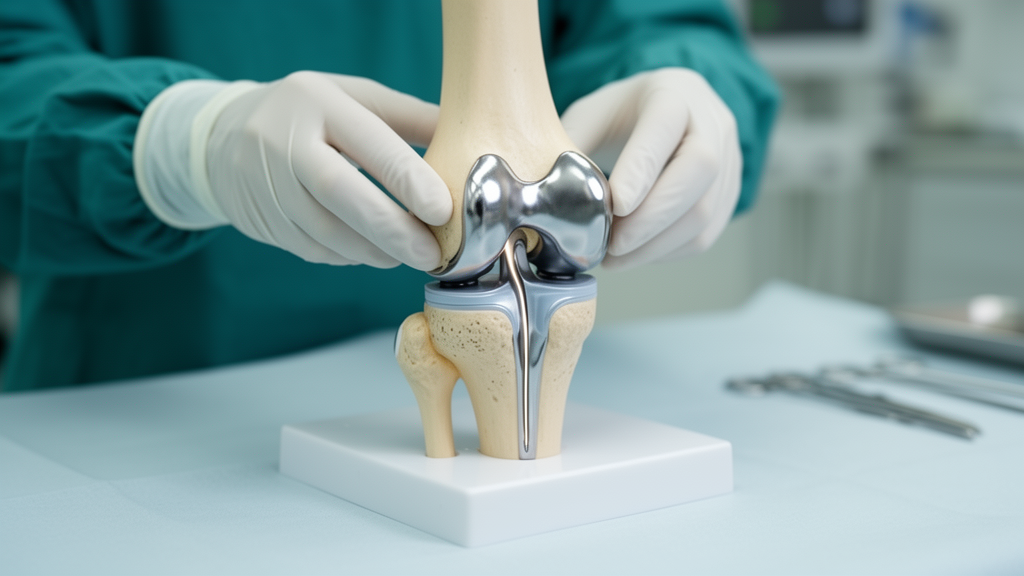
Patients with medial compartment knee osteoarthritis often show a subtle radiographic finding that has divided surgeons for years: on a standing front-view X-ray, the femur sits shifted slightly toward the inner side of the tibia. This displacement, called coronal tibiofemoral subluxation, has been treated by some as a warning sign that a partial knee replacement might fail. A new retrospective cohort study of 457 knees now reports that, at least over two years, the degree of this subluxation before surgery did not translate into worse clinical outcomes after a mobile-bearing medial unicompartmental knee arthroplasty, provided the deformity was correctable and standard selection criteria were met.
The study, published in Archives of Orthopaedic and Trauma Surgery, analyzed consecutive knees with anteromedial osteoarthritis that underwent surgery at a single center between August 2016 and May 2023. Anteromedial osteoarthritis is a specific pattern of medial compartment wear in which cartilage loss is concentrated at the front and middle of the tibial plateau, the lateral joint space is preserved, and the varus deformity can be passively corrected toward neutral alignment. All operations used the cementless Oxford Phase 3 mobile-bearing implant inserted through a minimally invasive medial parapatellar approach without patellar eversion, performed by two high-volume surgeons with consistent indications and techniques throughout the study period.
To quantify subluxation, the researchers measured the medial displacement of the lateral femoral condyle relative to the total width of the tibial plateau on preoperative weight-bearing anteroposterior radiographs, excluding osteophytes and measuring from true cortical margins. Expressed as a percentage of tibial plateau width, this ratio-based approach normalizes for individual knee size. Knees were then sorted into three predefined severity groups: mild subluxation from 0 to 5 percent, moderate from 5 to below 10 percent, and severe at 10 percent or more. Because no universally accepted thresholds exist for this measurement in partial knee replacement, the cut-offs were set before analysis to create clinically interpretable groups while keeping adequate sample sizes in each category.
The clinical outcome measure was the Oxford Knee Score, a patient-reported questionnaire collected prospectively as part of routine follow-up at four time points: before surgery and at 3 months, 1 year, and 2 years afterward. Follow-up rates ranged from 84.0 to 90.5 percent across these time points. The minimal clinically important difference for this score has been reported at approximately 5 points, giving the team a benchmark for judging whether any observed differences between groups would matter to patients. Radiographic measurements were performed without blinding to group allocation but were blinded to clinical outcomes, and reproducibility testing showed acceptable reliability, with an intraobserver intraclass correlation coefficient of 0.864 and an interobserver coefficient of 0.836.
The central result was strikingly uniform. At two years, mean improvements in the Oxford Knee Score were 16.6 plus or minus 8.9 points in the mild group, 14.8 plus or minus 8.3 in the moderate group, and 16.0 plus or minus 9.3 in the severe group. All three groups exceeded the reported minimal clinically important difference, while the differences between groups fell below it. When the researchers calculated adjusted mean differences controlling for age, sex, body mass index, and preoperative score, the between-group differences remained small, with confidence intervals that included zero, meaning no statistically reliable advantage or disadvantage for any severity category. A sensitivity analysis restricted to knees with complete two-year score data produced findings consistent with the primary analysis.
The study also examined whether subluxation severity left fingerprints on preoperative joint geometry or on the choices surgeons made in the operating room. The severe subluxation group showed a greater joint line convergence angle than both the mild and moderate groups, a difference that held in post-hoc testing with p values below 0.001, and subluxation severity correlated modestly with this angle at a Spearman coefficient of 0.29. Lower lateral distal femoral angle values appeared in the moderate and severe groups compared with the mild group. These patterns suggest that subluxation partly reflects underlying bone and joint-line morphology, particularly of the lateral femoral condyle, but the modest correlation strength points to limited clinical impact of this relationship on its own.
Bearing thickness, the thickness of the mobile polyethylene insert selected during surgery, served as a proxy for intraoperative ligament balancing. Prior work in total knee arthroplasty had suggested that pronounced subluxation can disrupt soft tissue balance and push surgeons toward thicker inserts. Here, mean bearing thicknesses were 3.6, 3.7, and 3.7 millimeters in the mild, moderate, and severe groups respectively, a non-significant difference with p equal to 0.428, and the correlation between subluxation severity and bearing thickness was negligible. The authors interpret this as evidence that preoperative coronal subluxation primarily reflects static joint morphology rather than true functional instability, and that restoring ligament balance during a mobile-bearing procedure may accommodate the preoperative displacement.
The mobile-bearing design is central to this interpretation. Because the floating bearing allows a degree of self-alignment and congruent articulation through the motion arc, the implant is thought to be less dependent on perfect preoperative joint geometry than a fixed-bearing construct, even though its lower constraint could theoretically make it more sensitive to ligament imbalance. In this cohort, that theoretical sensitivity did not materialize into measurable two-year harm. The authors conclude that within the setting of correctable deformity and appropriate patient selection, coronal tibiofemoral subluxation alone should not be considered a contraindication to mobile-bearing medial unicompartmental knee arthroplasty.
Important caveats frame how far this conclusion travels. The study was retrospective, included only knees whose varus deformity could be manually corrected to neutral on preoperative examination, and did not evaluate how subluxation behaved under valgus stress or whether it persisted after surgery. Postoperative radiographs were not assessed longitudinally, so residual subluxation and its potential to drive lateral compartment cartilage degeneration, a concern raised by earlier studies, remain unexamined here. Alignment was measured on short knee radiographs rather than full-length lower limb films, both knees of bilateral patients were included, missing questionnaire data were handled with available-case analysis, and knees requiring revision or reoperation within two years were excluded, which may introduce survivorship bias. All procedures were done by high-volume surgeons at one center, and only a mobile-bearing implant was tested, leaving generalizability to fixed-bearing designs uncertain. The relatively small number of knees at two-year follow-up in the severe group may also have raised the risk of a type II error.
Within those limits, the practical message is that a measurable degree of preoperative coronal subluxation, by itself, did not doom patients to inferior short-term results when the deformity was correctable and standard indications were satisfied. The authors note that measurement techniques for this subluxation vary across the literature and lack standardized anatomical landmarks, which complicates comparisons between studies, and they call for longer-term survivorship and radiographic progression analyses to determine whether the two-year equivalence holds as implants age. For now, the findings suggest surgeons weighing a partial knee replacement can place less weight on this single preoperative X-ray parameter, interpreting it instead alongside ligament balance and the presence of subluxation on lateral radiographs when making the final decision.
Subject of Research: The influence of preoperative coronal tibiofemoral subluxation on two-year outcomes after mobile-bearing medial unicompartmental knee arthroplasty for anteromedial osteoarthritis
Article Title: Preoperative coronal tibiofemoral subluxation does not affect 2-year outcomes after mobile-bearing medial unicompartmental knee arthroplasty for anteromedial osteoarthritis
Article References: Kinoshita, T., Bunyoz, K. I., Mortensen, K. R. L., Bredgaard Jensen, C., Price, A., Gromov, K., & Troelsen, A. (2026). Preoperative coronal tibiofemoral subluxation does not affect 2-year outcomes after mobile-bearing medial unicompartmental knee arthroplasty for anteromedial osteoarthritis. Archives of Orthopaedic and Trauma Surgery, 146(1), Article 387. https://doi.org/10.1007/s00402-026-06530-x
Image Credits: AI Generated
DOI: 10.1007/s00402-026-06530-x
Keywords: unicompartmental knee arthroplasty, coronal tibiofemoral subluxation, anteromedial osteoarthritis, Oxford Knee Score, mobile-bearing implant, knee osteoarthritis, radiographic measurement, joint line convergence angle, bearing thickness, retrospective cohort study, patient selection, two-year follow-up
News Source: Ophelia Keating. (October 29, 2026). Knee Subluxation Before Surgery Does Not Worsen Two-Year Results of Partial Knee Replacement. Scienmag.



