


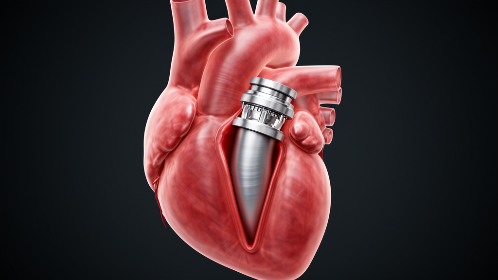
The case, published in General Thoracic and Cardiovascular Surgery Cases, concerns a 69-year-old woman who developed prosthetic valve dehiscence and a large aortic root pseudoaneurysm four months after aortic valve replacement for infective endocarditis. Because her native aortic annulus was completely destroyed, the team performed a redo Bentall procedure using the graft insertion technique, and postoperative imaging subsequently revealed a markedly narrowed outflow tract despite the implantation of an appropriately sized valve.
The graft insertion technique was introduced as a salvage approach to achieve secure proximal fixation in redo aortic root surgery when extensive annular destruction has occurred. Redo aortic root surgery after infective endocarditis complicated by annular destruction remains technically challenging. Extensive debridement is mandatory to eradicate infection, but once annular continuity is lost, secure proximal reconstruction becomes difficult and conventional techniques may fail. The graft insertion technique, originally described by Nakamura and colleagues, was introduced to achieve secure proximal fixation in exactly these complex situations, and recent reports including a midterm outcome study by Narita and colleagues have demonstrated favorable results with no recurrence of pseudoaneurysm or major aortic events.
The patient’s story began four months earlier, when she underwent aortic valve replacement with a 21-mm bioprosthetic INSPIRIS valve for infective endocarditis. Preoperative computed tomography performed before that initial operation documented her baseline left ventricular outflow tract geometry: a circumference of 83.2 mm, a maximum diameter of 30.9 mm, a minimum diameter of 21.7 mm, and an area of 5.18 square centimeters. Those figures matter because they show that her outflow tract was not markedly small to begin with, a point the authors emphasize when attributing the later narrowing primarily to the reconstruction itself rather than to unfavorable baseline anatomy.
During follow-up, abnormal mobility of the prosthetic valve was first detected on transthoracic echocardiography. Transesophageal echocardiography then demonstrated marked rocking motion of the valve, suggesting dehiscence, in which the prosthesis detaches from its anchoring ring. Contrast-enhanced computed tomography confirmed pseudoaneurysm formation at the annular regions of the left and right coronary cusps, with detachment of the prosthetic valve from the annulus. Surgical reintervention was indicated because of the risk of rupture and progressive prosthetic valve instability.
The operation itself did not go smoothly. Redo median sternotomy, reopening the chest through the previous incision, was attempted, but a major right ventricular injury occurred during reentry. Emergent cardiopulmonary bypass was established and the chest was safely reopened under circulatory support. Intraoperative examination revealed complete loss of the aortic wall at the left and right coronary cusp annular regions corresponding to the pseudoaneurysm cavities, but no macroscopic evidence of active infection. The authors note that the pseudoaneurysm cavity was surrounded by fibrotic tissue and the aortic wall had been replaced by fragile but noninfected tissue, suggesting the dehiscence was a consequence of previous infection rather than ongoing inflammatory destruction.
Because the annulus was extensively destroyed, the team selected the graft insertion technique. Intraoperative assessment showed that a 19-mm sizer could pass through the annular region whereas a 21-mm sizer could not, indicating an effective annular diameter between 19 and 21 mm. A 24-mm Valsalva vascular graft was inverted, using its straight tubular portion rather than the sinus segment, and inserted into the left ventricular cavity. Eleven pledgeted 2-0 braided polyester sutures were placed circumferentially from inside the left ventricle toward the epicardial side and tied securely, and the proximal anastomosis was reinforced with a continuous 4-0 polypropylene suture. Notably, a ball sizer or Hegar dilator was not used during knot tying, an omission the authors identify as a potentially important technical factor. The graft was then pulled out and anastomosed to a composite graft carrying a 19-mm bioprosthetic valve. During the procedure, injury to the right coronary artery required additional coronary artery bypass grafting with a saphenous vein graft, and the injured right ventricular wall was repaired with a bovine pericardial patch. Total operation time was 423 minutes, cardiopulmonary bypass time 315 minutes, and aortic cross-clamp time 189 minutes.
Even before the operation ended, the surgeons had concerns. During insertion of the inverted graft into the outflow tract, narrowing was visually suspected intraoperatively. Those suspicions were confirmed after surgery. Contrast-enhanced computed tomography demonstrated significant narrowing of the outflow tract at the level of the inverted graft, with a minimal diameter of 14 mm, despite the implantation of a 19-mm bioprosthetic valve. For comparison, the minimum diameter before the initial surgery had been 21.7 mm, so the reconstructed channel had lost roughly a third of its smallest dimension.
The functional consequences were then assessed with an imaging technique rarely applied to this problem. Transthoracic echocardiography was attempted first, but adequate visualization of the outflow tract was not achieved and Doppler-derived flow velocities could not be reliably obtained. The team therefore turned to four-dimensional flow magnetic resonance imaging, a modality that captures blood flow in three dimensions over the cardiac cycle. The scan demonstrated accelerated systolic flow at the reconstructed outflow tract and allowed quantitative assessment of flow velocity, indicating functional left ventricular outflow tract stenosis rather than merely an anatomical appearance. The authors argue that this combination of morphological and hemodynamic evaluation confirms the narrowing represented true functional stenosis rather than a transient or imaging-related artifact.
Why did the channel narrow when a 24-mm graft and a 19-mm valve had been used? The authors analyze several contributing mechanisms. First, the native outflow tract geometry may have provided limited tolerance to additional narrowing. Second, the intraventricular portion of the inverted graft itself may have reduced the lumen. However, they consider the most relevant mechanism to be the circumferential placement of pledgeted mattress sutures and reinforcement materials, including a donut-shaped felt strip and a bovine pericardial patch, at the proximal anastomosis. After graft inversion and pull-through, these materials are distributed around the circumference and effectively reduce the internal radius of the reconstructed outflow tract. Even with a 24-mm graft, the cumulative thickness of graft material, pledgets, and felt reinforcement can shrink the effective internal diameter substantially, producing a marked discrepancy between the expected and observed dimensions.
The clinical outcome was favorable: the postoperative course was uneventful and the patient was transferred to a rehabilitation facility without signs of recurrent infection. But the authors stress that the case exposes an important pitfall of the graft insertion technique. Even when infection has been eradicated and an appropriately sized prosthetic valve is used, outflow tract narrowing may occur as a consequence of the reconstruction itself. They recommend meticulous technical attention, including appropriate graft sizing, careful management of the proximal anastomosis, and the use of a ball sizer or Hegar dilator during knot tying to maintain luminal diameter. They also propose that postoperative multimodality imaging, particularly contrast-enhanced computed tomography and four-dimensional flow MRI, may play an essential role in detecting functional outflow tract stenosis and ensuring the hemodynamic safety of this technique in complex aortic root reconstruction. As a single case report, the observation does not establish how frequently such narrowing occurs after the graft insertion technique, an issue the authors note has received limited attention because most previous studies focused on infection control and anastomotic security rather than postoperative outflow tract geometry.
Subject of Research: Left ventricular outflow tract stenosis after redo Bentall surgery using the graft insertion technique
Article Title: Left ventricular outflow tract stenosis following Bentall procedure using the graft insertion technique: a case report
Article References: Yokawa, K., Yoshida, K., Ishimoto, K., Nakagawa, T., Higuma, T., Tanaka, Y., Oshima, Y., Obo, H., & Wakiyama, H. (2026). Left ventricular outflow tract stenosis following Bentall procedure using the graft insertion technique: a case report. General Thoracic and Cardiovascular Surgery Cases, 5(1), Article 37. https://doi.org/10.1186/s44215-026-00265-2
Image Credits: AI Generated
DOI: 10.1186/s44215-026-00265-2
Keywords: aortic root reconstruction, Bentall procedure, graft insertion technique, left ventricular outflow tract, infective endocarditis, prosthetic valve dehiscence, 4D-flow MRI, pseudoaneurysm, cardiac surgery, computed tomography, Left, ventricular
News Source: Ophelia Keating. (October 30, 2026). Inverted Graft Technique After Bentall Surgery Triggered Unexpected Outflow Tract Narrowing. Scienmag.



