

Brain tumors exhibit distinct growth behaviors that significantly influence patient outcomes and treatment approaches. Fundamentally, these tumors adopt one of two aggressive strategies to expand within the confined space of the skull: they either exert mechanical pressure by pushing against the surrounding brain tissue or infiltrate by sending invasive, finger-like projections that destroy neighboring neural structures. A deeper understanding of these divergent tactics is critical, not only because the mode of growth relates directly to neurological symptoms, but also because it offers new avenues for clinical intervention and prognostication.
Previous investigations have underscored that tumors which physically displace brain tissue through mechanical force often induce more pronounced neurological dysfunction than those that primarily destroy tissue. This disruption is thought to arise from the swelling and deformation that the brain undergoes as it contends with the solid forces exerted by the tumor mass. Untangling the complexities of how mechanical stress impacts the brain has posed challenges historically, limited by the difficulty of adequately measuring these forces in vivo during surgery and correlating them with clinical outcomes.
In a groundbreaking study published in the journal Clinical Cancer Research, a multidisciplinary team of researchers from the University of Notre Dame, Harvard Medical School, Massachusetts General Hospital, and Boston University have pioneered an innovative intraoperative technique that captures the mechanical stresses exerted by brain tumors. Utilizing advanced 3D neuronavigation technology widely available in neurosurgical suites, their method allows surgeons to quantify the pressure a tumor applies on brain tissue in real-time during tumor resection procedures. This measurement, they show, can differentiate between glioblastoma tumors—primary brain cancers known for their invasive nature—and metastatic tumors that originate elsewhere in the body and spread to the brain.
.adsslot_9SW0Ft8yJR{width:728px !important;height:90px !important;}
@media(max-width:1199px){ .adsslot_9SW0Ft8yJR{width:468px !important;height:60px !important;}
}
@media(max-width:767px){ .adsslot_9SW0Ft8yJR{width:320px !important;height:50px !important;}
}
ADVERTISEMENT
Central to this development is the marriage of biomechanical engineering with clinical neuroscience. Neurosurgeons conventionally rely on pathological assessment of tumor samples, where a tissue slice is reviewed live to identify tumor type and inform subsequent treatment pathways, such as chemotherapy or radiation regimens. The novel approach introduced by Meenal Datta, an assistant professor of aerospace and mechanical engineering at Notre Dame, enhances this workflow by adding a brief yet powerful step: measuring the solid mechanical stress during surgery helps classify tumors mechanically, independent of histological analysis.
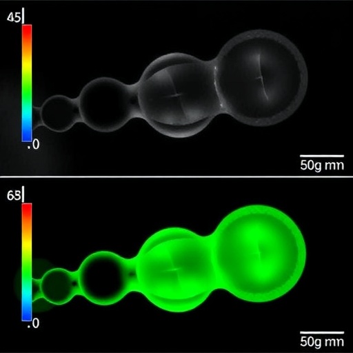
The method involves detailed assessment of patient-specific data, integrating preoperative magnetic resonance imaging (MRI) scans with intraoperative measures taken as the surgeon exposes the brain. The Brainlab neuronavigation platform serves as a pivotal tool that provides a dynamic 3D map, enabling precise identification of brain tissue displacement or “bulge” caused by tumor swelling prior to its resection. Through computational modeling and point-based measurement systems, the research team translates these visual and spatial data into quantitative estimates of the forces tumors exert and the corresponding amount of brain tissue lost.
The team’s findings elucidate a key biomechanical distinction: tumors that primarily cause displacement tend to generate substantial swelling in the adjacent brain, whereas those that replace brain tissue by invading and destroying it produce less swelling. This difference is critical for surgeons and oncologists as it refines the understanding of tumor behavior in vivo. It suggests that mechanical stress measurements are not mere supplemental data but essential biomarkers that could influence treatment decisions and symptom management.
Developing these computational models required sophisticated biomechanical simulations that account for tissue elasticity, tumor rigidity, and fluid dynamics within brain parenchyma. The researchers’ ability to capture these variables intraoperatively heralds a new era in personalized neurosurgical care, wherein doctors can more accurately gauge the tumor’s impact on cerebral physiology and plan interventions accordingly.
Funded by prominent bodies such as the National Institutes of Health and the U.S. National Science Foundation, this research bridges a critical gap between engineering principles and clinical oncology. It marks one of the earliest demonstrations that applying mechanical science to neuro-oncology can yield practical tools with direct implications for patient management. Notably, having precise knowledge of mechanical force exerted by tumors offers tangible benefits: it could guide the use of steroids to mitigate brain swelling or inform the administration of antipsychotic agents to counter neurocognitive side effects.
Moreover, this mechanical approach transcends tumor type, displaying a disease-agnostic quality that lauds its broader applicability. The research team employed animal models involving breast cancer brain metastases, glioblastomas, and pediatric ependymomas. Strikingly, in the breast cancer metastasis model, changes in tumor mechanical force predicted chemotherapy response earlier than conventional imaging detected tumor shrinkage. This suggests that biomechanical parameters might serve as sensitive early indicators of treatment efficacy, a crucial advancement given the limitations of current imaging modalities.
Looking ahead, Professor Datta and her colleagues envision that expanding the use of these mechanical force measurements will not only improve symptom control and therapeutic outcomes but also accelerate the integration of biomechanical insights into routine clinical practice. They advocate for further validation studies and broader adoption of intraoperative stress estimation as a tool to refine prognosis and tailor individualized therapeutic strategies.
Collaboration across engineering disciplines, neurosurgery, and oncology underpins this innovative work. Besides Datta, key contributors include Hadi T. Nia from Boston University, Ashwin S. Kumar from Massachusetts General Hospital and Harvard Medical School, and Saeed Siri from Notre Dame, among other notable experts. This collective expertise has culminated in a versatile methodology that leverages accessible technology, making it feasible for widespread clinical deployment without the need for specialized or prohibitively expensive equipment.
Alongside her role at Notre Dame’s aerospace and mechanical engineering department, Datta’s affiliations span several interdisciplinary research institutes focused on cancer, precision health, material science, and rare diseases. This broad institutional support has bolstered her capacity to blend mechanical engineering methods with biomedical applications, addressing a pressing unmet need in brain cancer treatment.
The implications of this research extend far beyond academic circles. For patients and their families, the ability to characterize tumors based on mechanical stress signifies hope for more accurate diagnoses, less invasive monitoring, and enhanced therapeutic precision. For clinicians, these insights could transform surgical planning and postoperative management, ultimately improving survival rates and quality of life for individuals grappling with devastating brain tumors.
As the medical community continues to unravel the complex interplay between tumor biology, brain mechanics, and patient outcomes, this pioneering study paves a promising path. It exemplifies how focused interdisciplinary innovation can yield revolutionary tools that meld the rigor of engineering with the intricacies of human health, charting the future course of neuro-oncological care.
Subject of Research: Brain Tumor Mechanics and Intraoperative Stress Measurement
Article Title: Solid Stress Estimations via Intraoperative 3D Navigation in Patients with Brain Tumors Open Access
News Publication Date: 1-Jul-2025
Web References:
https://aacrjournals.org/clincancerres/article/doi/10.1158/1078-0432.CCR-24-4159/762916/Solid-stress-estimations-via-intraoperative-3D
https://engineering.nd.edu/faculty/meenal-datta/
https://pubmed.ncbi.nlm.nih.gov/30948807/
Image Credits: University of Notre Dame/Wes Evard
Keywords: Brain cancer, Glioblastomas, Metastasis, Mechanical stress, Imaging
Tags: advancements in tumor growth researchbrain tumor growth patternsbrain tumor research studieschallenges in measuring mechanical stress in vivoclinical intervention strategies for brain tumorsinfiltration of brain tumorsmechanical pressure in brain tumorsmultidisciplinary approaches to brain cancerneurological symptoms of brain tumorspatient outcomes in brain tumor therapyprognostication in brain tumor treatmentunderstanding brain tumor behaviors


