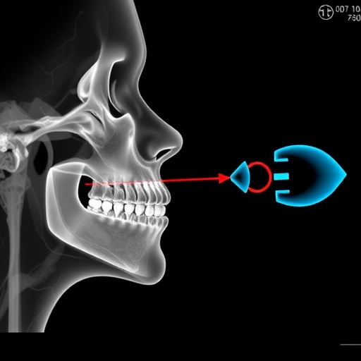
In a pioneering breakthrough, researchers at Stanford University, working alongside scientists from the German Cancer Institute, have unveiled a transformative approach in the visualization of injectable filler materials during injection laryngoplasty by utilizing shortwave infrared (SWIR) imaging technology. This innovative technique promises to revolutionize the precision and effectiveness of vocal fold augmentation procedures commonly performed to address vocal fold paralysis and various other forms of glottic insufficiency. The findings, detailed in the latest edition of Otolaryngology–Head and Neck Surgery, introduce a method that elevates the real-time visualization capabilities dramatically beyond current imaging standards.
Injection laryngoplasty is a delicate procedure where filler materials are injected into the vocal folds to improve voice function by increasing tissue bulk and enabling better vocal fold closure. Traditional visualization methods rely heavily on visible light scopes, which often face challenges in accurately distinguishing filler materials beneath the mucosal surface. Leveraging SWIR imaging, operating within the 1000–2000 nanometer spectrum, overcomes these hurdles by producing high-contrast images that crisply differentiate the injected material from surrounding tissues. This enhanced contrast arises largely due to variations in water content between vocal fold tissue and adjacent respiratory mucosa, particularly accentuated at a wavelength of 1550 nm.
The technical foundation of the SWIR imaging system employed by the researchers involved a multimodal approach combining both reflection and fluorescence imaging modes. Reflection imaging detects the light that bounces back from tissue structures, while fluorescence imaging captures emitted light after tissue excitation, providing complementary data sets that together render a comprehensive tissue characterization. This synergy unlocks the ability for surgeons to directly observe the precise placement of fillers in real-time, to identify any superficial misplacements immediately, and to monitor the gradual resorption or degradation of the injected substances over time.
The implications for surgical practice are profound. Real-time, high-contrast visualization during injection laryngoplasty mitigates the risk of inaccurate injections, which can lead to suboptimal vocal outcomes or complications. By enabling surgeons to verify treatment efficacy intraoperatively, SWIR imaging could considerably reduce the need for repeat procedures and enhance patient satisfaction. Furthermore, the technology’s capacity for fine differentiation of tissue layers offers a novel lens through which the dynamic interplay of biological structures can be studied during surgery, providing insights previously unattainable through conventional visualization techniques.
While the existing research was conducted ex vivo—meaning on extracted tissues outside of a live organism—the authors emphasize the practicality of integrating SWIR imaging hardware into standard endoscopic or operating microscope systems. Notably, the financial barriers to adoption have diminished due to significant reductions in the cost of SWIR cameras over recent years. This affordability positions the technology as an accessible upgrade for clinical centers, potentially setting a new standard for otolaryngological procedures involving soft tissue visualization.
Beyond the scope of injection laryngoplasty, this cutting-edge imaging modality exhibits promising applications across a broad spectrum of laryngeal disorders. For instance, its sensitivity to variations in water and collagen content could facilitate early detection and nuanced assessment of vocal fold cysts, polyps, and other pathological anomalies. Additionally, SWIR imaging’s ability to differentiate subtle inflammatory changes within the laryngeal mucosa may contribute significantly to the diagnosis and monitoring of chronic laryngeal inflammation, potentially improving intervention timing and outcomes.
Moreover, the multimodal SWIR platform could serve as a pivotal tool in research settings by enabling a detailed investigation into the biomechanical properties and physiological behaviors of vocal fold tissues. This could pave the way for new therapeutic protocols and enhanced understanding of voice disorders at a microstructural level. Its non-invasive nature and compatibility with real-time imaging make it particularly suited for longitudinal studies tracking tissue changes across treatment timelines.
The research team spearheading this innovation is led by Dr. Tulio A. Valdez, Professor of Otolaryngology – Head and Neck Surgery at Stanford Medicine, whose expertise and advocacy underscore the clinical relevance and transformative potential of SWIR imaging. Dr. Valdez articulates a vision wherein surgical accuracy and patient outcomes are significantly elevated, facilitated by high-resolution, high-contrast imagery that grants surgeons unprecedented visibility beneath the mucosal surface during intervention.
Integration into routine clinical workflows, however, must reconcile certain technical challenges, such as the development of miniaturized, user-friendly SWIR imaging devices compatible with delicate endoscopic instrumentation. Ongoing engineering efforts aim to optimize system robustness, image processing algorithms, and ergonomic deployment in surgical theatres to ensure seamless adoption by otolaryngologists worldwide.
The study’s publication in Otolaryngology–Head and Neck Surgery, the prestigious peer-reviewed journal of the American Academy of Otolaryngology–Head and Neck Surgery Foundation (AAO-HNS/F), ensures rigorous scientific validation and broad dissemination among clinical specialists. The AAO-HNS/F itself constitutes a leading authority in the field, representing over 13,000 experts dedicated to advancing head and neck surgical practices. Their endorsement signifies confidence in the clinical utility and innovation embodied by this SWIR imaging research.
With vocal fold paralysis and glottic insufficiency affecting countless patients globally, innovations like the application of shortwave infrared imaging represent a critical step forward in otolaryngology. By fundamentally enhancing the surgeon’s ability to visualize soft tissue interactions and injectable materials, this technological leap holds the promise of defining a new paradigm in precision-guided laryngeal surgery.
As SWIR imaging technology continues to mature and find its footing within clinical settings, future research and development will likely expand its applications further, integrating artificial intelligence-driven image analysis and augmented reality overlays. Such advancements could transform surgeries from visually guided procedures into data-enriched, precision interventions with unprecedented levels of accuracy and patient safety.
This research not only exemplifies the power of interdisciplinary collaboration between engineering and medical sciences but also highlights the ongoing commitment to improving quality of life for patients with vocal disorders. With clinical translation on the horizon, SWIR imaging offers a promising glimpse into the future of minimally invasive, highly precise treatment modalities that harness the unique properties of the infrared spectrum to unveil what lies beneath the surface.
Subject of Research: Visualization of injectable filler materials during injection laryngoplasty using shortwave infrared (SWIR) imaging.
Article Title: Multimodal Shortwave Infrared Imaging for Visualization of Injection Laryngoplasty.
News Publication Date: 17-Oct-2025.
Web References: https://aao-hnsfjournals.onlinelibrary.wiley.com/doi/10.1002/ohn.70050
References: Park, R.K., Lee, M.C., Härtl, S., Arús, B.A., Nuyen, B., Sung, C.-K., Baik, F.M., Bruns, O.T. and Valdez, T.A. (2026), Multimodal Shortwave Infrared Imaging for Visualization of Injection Laryngoplasty. Otolaryngol Head Neck Surg, 174: 185-194.
Keywords: Otolaryngology, Injection Laryngoplasty, Shortwave Infrared Imaging, SWIR, Vocal Fold Paralysis, Glottic Insufficiency, Real-time Imaging, Multimodal Imaging, Fluorescence Imaging, Reflection Imaging, Laryngeal Surgery, Vocal Fold Augmentation.
Tags: 1550 nm wavelength for medical imagingadvanced imaging in glottic insufficiency treatmentenhanced contrast imaging in otolaryngologyinjectable filler differentiation using SWIRovercoming visible light limitations in vocal fold proceduresprecision imaging for vocal fold paralysis treatmentreal-time visualization in vocal fold augmentationshortwave infrared imaging in vocal fold injectionStanford University vocal fold researchSWIR technology for injection laryngoplastyvocal fold injection accuracy improvementvocal fold tissue imaging techniques


