Xu, Yang, and Zhong’s review in Frontiers in Cellular and Infection Microbiology evaluates the diagnostic performance, biological rationale, and evidence directness of conventional and emerging inflammatory biomarkers in pyogenic spinal infection. The authors conclude that circulating inflammatory markers cannot function as standalone diagnostic tests for pyogenic spinal infection, but are instead best interpreted within an evidence-informed clinical framework that systematically synthesizes patient presentation, magnetic resonance imaging, microbiological confirmation, and longitudinal host inflammatory kinetics. Pyogenic spinal infection, which encompasses pyogenic vertebral osteomyelitis and infectious spondylodiscitis, remains notoriously difficult to identify at an early stage because initial clinical manifestations are frequently nonspecific, imaging findings overlap with degenerative or postoperative spinal pathologies, and definitive microbiological identification is often delayed or unavailable. Consequently, diagnostic uncertainty does not stem from the inadequacy of any single laboratory assay, but rather from the clinical reality that radiological, microbiological, and inflammatory signals emerge asynchronously during the disease course.
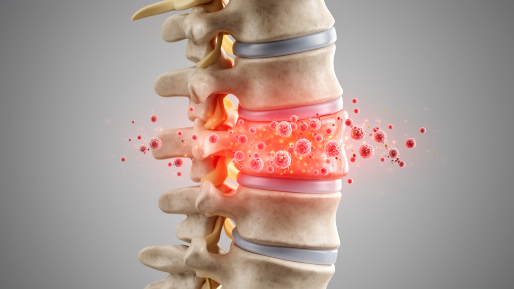
The clinical presentation of pyogenic spinal infection is characterized by notable heterogeneity and an insidious onset, with persistent focal neck or back pain representing the predominant early symptom. Local tenderness, paraspinal muscle spasms, and restricted spinal motion frequently occur, whereas classic systemic signs such as fever, chills, and malaise are inconsistently present or completely absent in early stages. As the infection progresses within the vertebral bodies, intervertebral discs, paraspinal tissues, or epidural space, expanding inflammatory masses or epidural abscesses may compress neural elements, causing radicular deficits, paresis, or sphincter dysfunction. Although spinal infection remains relatively rare, its reported incidence has steadily climbed in recent years, driven by population aging, a growing burden of chronic comorbidities such as diabetes and renal failure, expanded use of immunosuppressive drugs, and the increasing frequency of invasive spinal procedures. Hematogenous seeding represents the predominant route of inoculation, followed by direct surgical contamination and contiguous spread, with Staphylococcus aureus remaining the leading causative pathogen across clinical cohorts.
Current clinical pathways for diagnosing pyogenic spinal infection rely fundamentally on magnetic resonance imaging, microbiological cultures, histopathological biopsy, and conventional inflammatory laboratory panels. Magnetic resonance imaging offers high sensitivity for detecting vertebral osteomyelitis, disc involvement, and epidural abscesses, but its specificity is constrained by overlapping radiological features observed in degenerative disc disease, acute vertebral fractures, malignancy, and normal postoperative healing. Microbiological culture remains the cornerstone of pathogen identification, yet its diagnostic yield varies widely depending on sampling accuracy, collected specimen volume, previous antimicrobial exposure, and whether liquid aspirates are retrieved during image-guided biopsy. Large clinical cohorts of native vertebral osteomyelitis demonstrate that while combining blood cultures with tissue sampling enhances overall pathogen recovery, a notable proportion of patients still remain culture-negative. Under such circumstances, a negative microbiological culture cannot be interpreted as definitive evidence against spinal infection when clinical suspicion and radiological features remain compelling.
In routine clinical practice, conventional inflammatory markers such as C-reactive protein, erythrocyte sedimentation rate, and peripheral white blood cell count serve as the primary laboratory tools for initial evaluation and ongoing surveillance. C-reactive protein is widely utilized due to its low cost, rapid availability, and sensitivity to acute inflammatory stimuli, allowing clinicians to gauge active inflammatory responses and monitor therapeutic progress. However, C-reactive protein lacks etiological specificity, rising substantially following surgical trauma, accidental injury, or autoimmune flare-ups, while frequently remaining normal or only minimally elevated during early native infection or in immunocompromised individuals. Similarly, erythrocyte sedimentation rate responds sluggishly to clinical changes, frequently lingering at elevated levels long after successful treatment, and is influenced by non-infectious confounders including anemia, advanced age, and chronic disease. Peripheral white blood cell counts demonstrate limited utility, as overt leukocytosis is often absent in localized, deep-seated, or chronic infections, meaning a normal leukocyte count cannot exclude spinal infection.
From a mechanistic perspective, bacterial invasion initiates an innate host response through the recognition of pathogen-associated molecular patterns, such as staphylococcal lipoteichoic acid and peptidoglycan, by pattern-recognition receptors including Toll-like receptor 2 and nucleotide-binding oligomerization domain-containing protein 2. Downstream activation of nuclear factor-κB, mitogen-activated protein kinases, and Janus kinase-signal transducer pathways stimulates the rapid release of early pro-inflammatory cytokines, specifically interleukin-1β, tumor necrosis factor-α, and interleukin-6. Because interleukin-6 concentrations often rise earlier than C-reactive protein, it has attracted attention as an early warning indicator. Nevertheless, discriminatory interleukin-6 cut-offs, such as reported thresholds between 15 and 20 picograms per milliliter, have been derived almost exclusively from postoperative spinal surgical site infection cohorts and cannot be extrapolated directly to native hematogenous or chronic pyogenic spinal infections, while circulating tumor necrosis factor-α remains restricted to exploratory research.
The review critically analyzes emerging candidate biomarkers, emphasizing the importance of distinguishing direct spinal infection data from evidence extrapolated from periprosthetic joint infection, sepsis, and non-spinal disorders. Procalcitonin correlates strongly with systemic bacterial dissemination and persistent bacteremic risk, but its concentrations generally remain low in localized, indolent spinal infections or following uncomplicated spinal surgery. Consequently, while elevated procalcitonin indicates systemic bacterial involvement, a low measurement does not rule out localized pyogenic spinal infection. Pentraxin 3, produced locally by neutrophils, vascular endothelial cells, and myeloid lineage cells, reflects tissue-level innate immune activation rather than systemic hepatic synthesis, offering potential utility when surgical trauma confounds C-reactive protein interpretation; however, direct validation in spinal cohorts remains scarce. Similarly, S100A8/A9, or calprotectin, represents sustained myeloid activation and neutrophil recruitment, but supporting evidence is largely derived from synovial fluid in periprosthetic joint infection or systemic sepsis cohorts like the prospective CASCADE study, leaving its diagnostic utility in pyogenic spinal infection unvalidated.
Differential diagnosis presents another major hurdle, as atypical organisms elicit variable inflammatory responses that overlap biochemically and clinically with pyogenic bacteria. Comparative studies demonstrate that while patients with pyogenic spondylodiscitis frequently exhibit higher leukocyte counts, erythrocyte sedimentation rates, and C-reactive protein levels than those with spinal tuberculosis or brucellosis, substantial overlap precludes establishing pathogen-specific numerical cut-offs. In spinal tuberculosis cohorts, multivariable analysis demonstrates that thoracic anatomical involvement and extensive bony destruction, rather than inflammatory marker magnitudes, independently predict tuberculous disease. Brucellar spondylodiscitis often presents with sweating, back pain, and rural or dairy exposure, requiring dedicated serology and culture rather than relying on modest inflammatory changes. Furthermore, fungal spinal infections, such as Candida spondylodiscitis, and rare conditions caused by Nocardia or nontuberculous mycobacteria, frequently present with normal leukocyte counts and indolent biomarker trajectories, demonstrating that mild peripheral inflammatory activity must never be interpreted as evidence against active infectious disease when imaging remains abnormal.
Because pyogenic spinal infection is anatomically confined to specific spinal compartments, systemic blood markers frequently fail to capture the local microenvironmental reality of deep-seated disease. In patients with localized lesions, prior antimicrobial exposure, or altered host immunity, peripheral blood indices may remain unremarkable despite striking radiological abnormalities. Local specimens, including paravertebral abscess fluid, intervertebral disc aspirates, epidural exudates, and intraoperative tissues, reside directly adjacent to the infectious focus and harbor concentrated granulocytic, myeloid, and tissue-degradation mediators. However, obtaining local specimens requires invasive procedures, such as image-guided needle aspiration or open surgical biopsy, which carry procedural risks of neural injury and hemorrhage. Studies comparing sampling techniques reveal that open surgical biopsy yields higher microbiological positivity rates than percutaneous fine-needle or core needle techniques. The review emphasizes that invasive sampling should be undertaken judiciously for specific clinical indications, such as pathogen isolation or clarifying discordant clinical-radiological findings, rather than routinely performed simply to measure local biomarker concentrations.
In longitudinal patient management, monitoring inflammatory trajectories over time provides substantially greater diagnostic and prognostic value than relying on isolated single-point biomarker measurements. In post-surgical settings, C-reactive protein typically follows a predictable temporal curve, and failure to decline, a prolonged plateau, or a secondary rebound serves as an early signal of complication. In large longitudinal cohorts, including investigations of over seven hundred patients with pyogenic vertebral osteomyelitis, C-reactive protein time-series modeling significantly improved recurrence prediction compared to static threshold evaluations. Crucially, discordance between laboratory trajectories and the clinical picture demands prioritized reassessment: persistent pain, escalating neurological impairment, or progressive radiological destruction despite declining blood markers warrants immediate investigation rather than false reassurance. Conversely, when clinical symptoms resolve, neurological examination remains stable, and inflammatory markers exhibit sustained downward trajectories, established guidelines advise against routine repeat magnetic resonance imaging, thereby preventing unnecessary clinical interventions and excessive healthcare utilization.
Looking toward future diagnostic paradigms, metagenomic next-generation sequencing and artificial intelligence represent promising adjunctive technologies, provided their technical limitations are recognized. Metagenomic sequencing broadens pathogen detection without requiring organism-specific primers, demonstrating detection rates above eighty-four percent in retrospective spinal cohorts where conventional culture achieved roughly forty-one percent. Nevertheless, metagenomic assays identify microbial nucleic acids rather than viable organisms, making them susceptible to environmental contamination, colonization, and background noise, while failing to provide phenotypic antimicrobial susceptibility. Concurrently, machine-learning models offer theoretical advantages in harmonizing multimodal clinical, radiological, and kinetic biomarker datasets to predict complications or treatment failure. However, existing artificial intelligence models in spinal infection remain predominantly retrospective, single-center, and lacking prospective external validation. The authors emphasize that future clinical deployment of computational tools requires rigorous adherence to standardized reporting frameworks such as TRIPOD-AI and PROBAST-AI, ensuring that emerging technologies genuinely augment, rather than replace, clinical multidisciplinary judgment.
Subject of Research: Diagnostic utility and evidence-graded integration of inflammatory biomarkers in pyogenic spinal infection
Article Title: Inflammatory biomarkers in pyogenic spinal infection: evidence, limitations, and integrated diagnostic perspectives
Article References: Xu, J., Yang, Y., & Zhong, W. (2026). Inflammatory biomarkers in pyogenic spinal infection: evidence, limitations, and integrated diagnostic perspectives. Frontiers in Cellular and Infection Microbiology, 16, Article 1863268. https://doi.org/10.3389/fcimb.2026.1863268
Image Credits: AI Generated
DOI: 10.3389/fcimb.2026.1863268
Keywords: pyogenic spinal infection, vertebral osteomyelitis, inflammatory biomarkers, C-reactive protein, procalcitonin, interleukin-6, metagenomic next-generation sequencing, artificial intelligence, Inflammatory, biomarkers, pyogenic, spinal
News Source: Kristina Jarvis. (November 12, 2026). Inflammatory Biomarkers in Pyogenic Spinal Infection Require Integrated Reassessment Framework. Scienmag.