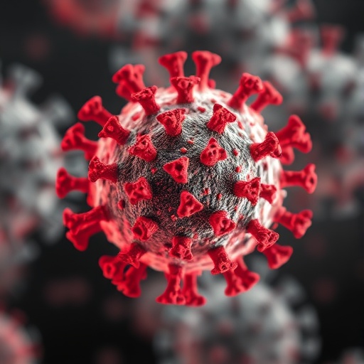
Scientists in India have shown that a single, largely overlooked structural protein of SARS-CoV-2 can, entirely on its own, assemble into virus-like particles that mount a substantial immune response in mice. The new study, published in Virology Journal, focuses on the membrane protein, or M protein, the most abundant component of the coronavirus envelope and the molecular scaffold that organizes the particle’s assembly. While most vaccine research has concentrated on the notorious spike protein, this work demonstrates that the membrane protein can self-assemble into round, virus-like particles roughly 180 to 200 nanometers in diameter and can drive both antibody and cellular immune responses in animals.
The team, led by Akash Kumar and Deepak Sehgal of the Shiv Nadar Institution of Eminence, with collaborators at the All India Institute of Medical Sciences, the University of Pittsburgh School of Medicine and King Saud University, began with computational work rather than wet-lab experiments. Using AlphaFold to predict the three-dimensional structure of the membrane protein from the Delta variant, refined through the YASARA energy-minimization server, they docked two copies of the protein together with ClusPro and analyzed the resulting dimer interface with PDBSum. The interaction analysis revealed one salt bridge, seven hydrogen bonds and 215 non-bonded van der Waals contacts holding the homodimer together. Four residues emerged as particularly important: glutamate 115, tyrosine 39, lysine 50 and glutamate 135. Glutamate 115 sits in the hinge region that governs the switch between the long and short conformations of the membrane protein dimer, a transition that cryo-electron microscopy studies had previously shown to be essential for virus assembly. Glutamate 135 lies in the C-terminal tail, where acidic residues help stabilize the intermolecular contacts needed for higher-order particle structure.
Molecular dynamics simulations then put the docked dimer through its paces. The team ran a 500-nanosecond simulation at 300 Kelvin and 1 bar using the OPLS force field in Desmond, tracking root-mean-square deviation, residue fluctuations, radius of gyration and solvent-accessible surface area. The results painted a picture of a rigid core with flexible edges: the C-alpha RMSD rose sharply at first, then plateaued at a stable value of roughly 8 to 9 angstroms, indicating that the complex relaxed into a new but durable conformation. The radius of gyration decreased and stabilized, and the solvent-accessible surface area declined, both signs that the dimer was packing more tightly over time. Only the peripheral helices and terminal tails showed meaningful mobility, consistent with the idea that membrane protein dimers act as stiff scaffolds that impose curvature on the viral membrane and provide a platform onto which the spike, envelope and nucleocapsid proteins are organized.
With the computational groundwork laid, the researchers turned to the baculovirus expression vector system, a workhorse of industrial protein production. They cloned the membrane gene into a recombinant bacmid and transfected Sf-21 insect cells, generating a P0 viral stock that was amplified and used to infect 500 million cells at a multiplicity of infection of 10. After 96 hours of incubation at 27 degrees Celsius, the cells were lysed with a Dounce homogenizer and sonication, and both the lysate and the concentrated culture medium were loaded onto a 20 to 80 percent sucrose gradient for ultracentrifugation at 27,000 rpm in an SW41 Ti rotor. A visible ring appeared at the junction of the 40 and 50 percent sucrose layers, corresponding to a particle density of 1.16 to 1.18 grams per milliliter, closely matching the density reported for purified SARS-CoV-2 virus-like particles and infectious coronavirions in earlier studies. SDS-PAGE and Western blotting confirmed the presence of the roughly 25-kilodalton membrane protein in these fractions, while uninfected Sf-21 cell controls produced no comparable structures.
The biophysical characterization confirmed that these were genuine, discrete particles rather than protein aggregates. Dynamic light scattering on a Horiba Zeta Sizer measured a hydrodynamic diameter of approximately 225 nanometers and a zeta potential of around minus 15 to minus 25 millivolts, the negative surface charge typical of enveloped virus particles. Field-emission scanning electron microscopy, at 40,000-fold magnification, revealed spherical to slightly pleomorphic particles of roughly 180 to 200 nanometers, and transmission electron microscopy of negatively stained samples showed numerous electron-dense spherical particles of about 200 nanometers embedded in a background of amorphous material. Taken together, the DLS, FESEM, TEM and immunoblotting data support the conclusion that expression of the membrane protein alone is sufficient to drive the budding of virus-like particles in this system, a capability that had not previously been demonstrated for the SARS-CoV-2 membrane protein in isolation.
The immunological evaluation was carried out in female BALB/c mice, aged four to six weeks, under protocols approved by the Institutional Animal Ethics Committee of Rodent Research India and compliant with Indian CCSEA standards. Each mouse received 100 micrograms of purified particles subcutaneously with 0.5 percent aluminum hydroxide adjuvant, while control animals received sterile phosphate-buffered saline. Blood samples collected at days 7, 14, 35 and 45 told a clear story. Serum IgG in the immunized group was already elevated by day 7, remained above control levels at all subsequent time points, and showed a gradual decline at days 35 and 45, a pattern consistent with a strong primary immune response rather than a loss of immunological memory. IgM peaked around day 7 and then declined but stayed above control levels through day 45. IgA, typically associated with mucosal immunity, was higher in the immunized group at every time point, indicating that the particles could stimulate class-switched antibody responses in addition to the early IgM wave. Isotype profiling on the final serum collection showed a broad antibody repertoire, with measurable IgG subclasses, strong IgM reactivity, and robust signals for kappa light chains and total heavy-plus-light chains, confirming active production of functional immunoglobulins.
The cellular arm of immunity proved equally responsive. Sandwich ELISA measured serum interferon-gamma, which was consistently higher in the immunized group with peak levels around day 14 and sustained elevation at days 35 and 45, a signature of T helper type 1 activation that mirrors findings from earlier SARS-CoV and SARS-CoV-2 structural protein vaccine studies. Quantitative real-time PCR on splenocytes deepened this picture. Using the 2^-ΔΔCt method on TRIzol-extracted RNA converted to cDNA, the team found markedly elevated expression of interferon-gamma, interleukin-2 and interleukin-12, together indicating a dominant Th1 pattern of the kind observed in SARS-CoV-2-specific T cells during infection and vaccination. Importantly, the particles also raised interleukin-4 and interleukin-13, showing that Th2 responses supporting antibody class switching were engaged as well, and a modest rise in interleukin-10 suggested a regulatory brake that could prevent excessive inflammation while preserving protective immunity. Th17 and transforming growth factor beta components rounded out a balanced cytokine profile. Statistical significance was assessed with two-way ANOVA and Tukey’s post-hoc test for the ELISA data and Mann-Whitney tests for the qRT-PCR data.
The question of neutralization, however, received a sober answer. Because the membrane protein has only a short N-terminal ectodomain and sits mostly embedded in the viral envelope, antibodies directed against it are not expected to block viral attachment or entry, and indeed the M-protein particles alone did not induce detectable neutralizing activity. This distinguishes them sharply from spike-targeted vaccines, which elicit the receptor-blocking antibodies that prevent infection. Yet the team argues that this is not a disqualifying limitation. The membrane protein has remained highly conserved across the many SARS-CoV-2 variants that have emerged since the pandemic began, including the Delta variant that originated in India, whereas spike has mutated relentlessly under immune pressure. A vaccine component built on the membrane protein could therefore provide durable immune memory and T-cell help that remains effective regardless of how the spike evolves, potentially serving as a conserved backbone in combination with variant-matched spike antigens. The researchers’ previous work on Membrane-Envelope particles had already shown antigenicity and neutralization activity, and co-expression of M and E proteins is known to enhance virion assembly and produce particles more faithful to the native virus, suggesting that head-to-head comparisons of M-only versus M-plus-E particles in larger animal cohorts are a natural next step.
The study also charts a path for refining the platform itself. Targeted mutations at glutamate 115 or glutamate 135 could probe how changes at the dimer interface affect particle formation, stability and immunogenicity, while tuning the lipid composition of the production system to favor lipids such as ceramide-1-phosphate, which is known to stabilize assembly-competent conformations of the membrane protein, might yield higher-quality particles. More definitive structural work, including immunogold labeling, cryo-electron microscopy and cryo-electron tomography, will be needed to visualize exactly how the membrane protein is oriented within the assembled particles. And before any translational claims can be made, neutralization assays, virus challenge studies and protection-efficacy experiments will be essential. For now, the finding stands as a striking demonstration of molecular self-assembly: one small, 25-kilodalton protein, acting alone in insect cells, can build a virus-like shell that the mouse immune system recognizes and attacks with vigor, opening a new avenue in the search for coronavirus vaccines built not on the shifting spike, but on the stable scaffold beneath it.
Subject of Research: Generation and immunological evaluation of SARS-CoV-2 membrane protein virus-like particles
Subject of Research: Biology
Article Title: Generation and immunological evaluation of SARS-CoV-2 membrane protein virus-like particles
Article References: Kumar, A., Inampudi, K. K., Kumar, V., Singh, R., Sinha, G. P., Parvez, M. K., & Sehgal, D. (2026). Generation and immunological evaluation of SARS-CoV-2 membrane protein virus-like particles. Virology Journal, 23(1), Article 185. https://doi.org/10.1186/s12985-026-03256-5
Image Credits: AI Generated
DOI: 10.1186/s12985-026-03256-5
Keywords: SARS-CoV-2, Membrane protein, Virus-like particles, VLPs, Immune response, TEM, BALB/c mice, Th1 response, Virology Journal
Cite Scienmag News
APA MLA Chicago
Kristina Jarvis. (September 10, 2026). Immunological evaluation of SARS-CoV-2 membrane protein virus-like particles. Scienmag. https://scienmag.com/immunological-evaluation-of-sars-cov-2-membrane-protein-virus-like-particles/
Kristina Jarvis. “Immunological evaluation of SARS-CoV-2 membrane protein virus-like particles.” Scienmag, 10 September 2026, https://scienmag.com/immunological-evaluation-of-sars-cov-2-membrane-protein-virus-like-particles/. Accessed 10 September 2026.
Kristina Jarvis. “Immunological evaluation of SARS-CoV-2 membrane protein virus-like particles.” Scienmag. September 10, 2026. https://scienmag.com/immunological-evaluation-of-sars-cov-2-membrane-protein-virus-like-particles/
Copy citation Download RIS
Tags: AlphaFold protein structure predictionAlphaFold structural predictionantibody and cellular immunity against SARS-CoV-2computational modeling of viral proteinscomputational virology researchcoronavirus envelope proteincoronavirus structural proteinsCOVID-19 vaccine developmentimmune response in miceimmune response to coronavirus structural proteinsimmunogenicity of coronavirus proteinsmembrane protein self-assemblynovel coronavirus vaccine targetsSARS-CoV-2 membrane proteinstructural analysis of viral protein interactionsvaccine development targetsvirus assembly mechanismsvirus envelope proteinvirus-like particle assemblyvirus-like particle characterizationvirus-like particle immune responsevirus-like particle immunogenicity