


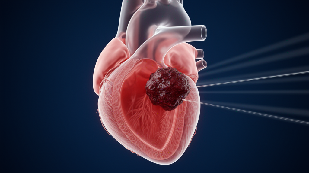
Doctors in Weifang, China, have reported an unusually detailed account of a rare and dangerous complication of an inherited heart-muscle disease: multiple blood clots lodged at the apex of the right ventricle of a 37-year-old woman with arrhythmogenic cardiomyopathy. The case, published in Frontiers in Cardiovascular Medicine, documents how two complementary imaging techniques—transthoracic echocardiography and cardiac magnetic resonance—were combined to reach a diagnosis that neither modality alone could have secured, and how six months of anticoagulant therapy led to complete resolution of the thrombi on follow-up imaging.
Arrhythmogenic cardiomyopathy is a rare inherited disorder in which heart-muscle tissue is progressively replaced by fibrous and fatty tissue. According to the 2020 Padua Criteria cited by the authors, the disease is now classified into right-ventricular-predominant, biventricular, and left-ventricular-predominant subtypes, with substantial variation in clinical presentation. Early on, patients may have no obvious symptoms; some first present with palpitations or syncope, and in advanced stages the disease can progress to right-heart or biventricular failure. Thrombus formation complicating the disease is uncommon—the authors state that the incidence reported in the existing literature is below five percent—but clots that detach can cause pulmonary or systemic embolism, sharply raising clinical risk.
The patient arrived at the hospital on September 10, 2025, complaining of recurrent chest tightness and shortness of breath that had begun more than three months earlier and worsened over the preceding half month. Her symptoms intensified with physical activity and did not ease with rest, and she also experienced intermittent nocturnal breathing difficulties. Physical examination found the apex beat displaced leftward and downward, with no pathological murmurs. Notably, her family history offered no clues: neither her parents nor other available relatives had any history of arrhythmogenic cardiomyopathy, unexplained arrhythmia, or sudden death, suggesting a sporadic rather than inherited origin in this instance.
Laboratory results pointed toward cardiac dysfunction. Her hemoglobin of 105 g/L indicated mild anemia, her uric acid of 432 μmol/L indicated hyperuricemia, and her NT-proBNP of 680 pg/mL suggested impaired cardiac function, while her D-dimer was 0.35 mg/L. The electrocardiogram showed sinus rhythm with complete right bundle branch block—a QRS duration of at least 120 milliseconds with an rSR’ pattern in leads V1–V2—along with marked right-axis deviation, ST-segment depression and T-wave inversion in leads V1–V3, premature ventricular complexes, and a prolonged QT interval. The authors note that this combination may suggest elevated right-ventricular pressure load. Twenty-four-hour Holter monitoring detected no non-sustained or sustained ventricular tachycardia, and the patient had no prior syncope. During exercise testing her breath-holding symptom worsened slightly and paired premature ventricular complexes appeared, findings the authors interpreted as possibly indicating elevated arrhythmic risk.
The decisive imaging began with transthoracic echocardiography. It measured a left-ventricular end-diastolic diameter of 40.1 mm, a left-ventricular ejection fraction of roughly 40 percent, a fractional shortening of about 23 percent, a right-ventricular transverse diameter of approximately 33.6 mm, a right-atrial transverse diameter of about 56.0 mm, a tricuspid annular systolic velocity of roughly 7 cm/s, a right-ventricular fractional area change of about 33 percent, and a tricuspid annular plane systolic excursion of about 16 mm. At the right-ventricular apex, the scan revealed diffuse myocardial trabeculation containing two clearly bounded, wall-attached hyperechoic masses measuring approximately 14.4 by 12.2 mm and 9.6 by 9.9 mm, which did not move significantly with the heartbeat. Moderate eccentric tricuspid regurgitation was also detected, with a peak flow rate of 2.36 m/s and a peak gradient of 22 mmHg. Chest computed tomography had already shown cardiomegaly, pericardial thickening, and a small pericardial effusion.
Cardiac magnetic resonance, performed two days after admission, consolidated the diagnosis. The right-ventricular end-diastolic volume indexed to body surface area was 121.94 mL/m², the left-ventricular indexed end-diastolic volume was 86.71 mL/m², and the right-ventricular ejection fraction was just 14 percent. The right-ventricular end-diastolic transverse diameter measured approximately 61.4 mm, with thinning and reduced motion of the apical wall. Cine imaging showed focal systolic wrinkling of the right-ventricular free wall in an accordion-like configuration and a low-signal lesion at the apex. Late gadolinium enhancement showed no delayed enhancement of the right-ventricular wall, and the apical mass was largely unenhanced with only a small area of internal enhancement—potentially indicative of porous thrombus tissue. Diffuse epicardial enhancement was also seen in the anterior and anterolateral walls of the left ventricle. Integrating all findings, the team established a diagnosis of right-ventricular-predominant arrhythmogenic cardiomyopathy with biventricular involvement, along with multiple mural thrombi at the right-ventricular apex, chronic heart failure, mild anemia, and hyperuricemia.
The authors argue that the two modalities play distinct and complementary roles. Echocardiography, as the preferred non-invasive first-line method, directly displays the diffuse apical trabeculation characteristic of the disease and identifies the size and attachment site of thrombi, providing early morphological evidence. They cite studies showing that echocardiographic parameters carry prognostic weight in this disease: a right-ventricular fractional area change below 23 percent is reported as the strongest predictor of major adverse cardiovascular events, while a tricuspid annular plane systolic excursion below 17 mm and right-ventricular dilation are also associated with increased risk. Cardiac magnetic resonance, meanwhile, establishes the diagnosis by demonstrating ventricular dilation, segmental wall-motion abnormalities, and characteristic late gadolinium enhancement, offering a comprehensive assessment of myocardial pathology. The authors also address an apparent discrepancy: the markedly different right-ventricular diameter measurements between the two techniques reflect inherent differences in imaging principles, acquisition planes, and scanning protocols, and semi-quantitative two-dimensional echocardiographic diameters should not be compared one-to-one with quantitative magnetic resonance values.
Distinguishing the apical mass from alternatives mattered clinically. The authors explain that trabecular hyperplasia shares signal intensity with normal myocardium and typically shows myocardial-like enhancement on late gadolinium sequences, whereas most cardiac tumors show early perfusion or enhancement, with malignant lesions tending toward prominent enhancement—features that help separate them from thrombus. They further distinguish arrhythmogenic cardiomyopathy from Brugada syndrome, which can be confused because both affect the right precordial leads: Brugada is primarily an ion-channelopathy with generally normal cardiac structure and a predisposition to polymorphic ventricular tachycardia or ventricular fibrillation, while arrhythmogenic cardiomyopathy involves fibrofatty replacement and structural remodeling, with relatively fixed T-wave inversions, epsilon waves, terminal conduction delay, and frequently monomorphic ventricular tachycardia.
On the mechanism of the clots, the authors speculate by reference to Virchow’s triad—blood stasis, endothelial injury, and hypercoagulability. Fibrofatty replacement of the apical myocardium may thin the wall and cause regional hypokinesia; diffuse trabeculation creates abnormal blood-flow pathways that reduce local flow velocity and promote platelet aggregation and fibrin deposition; and the fibrofatty lesions damage the endocardium, exposing collagen and activating the intrinsic coagulation pathway, with trabecular hypertrophy adding local frictional injury. Severe left-ventricular dysfunction and mild anemia may have further promoted thrombosis through hemodynamic disturbance. While single right-ventricular apical thrombi are relatively well described, the authors note that multiple thrombi confined exclusively to the apex, as in this patient, are comparatively uncommon.
Treatment combined rivaroxaban 20 mg once daily for in-hospital anticoagulation with supportive therapy: spironolactone to reduce cardiac load, sacubitril-valsartan and sustained-release metoprolol succinate to improve cardiac function and delay remodeling, folic acid and iron for the anemia, and benzbromarone to lower uric acid. The patient’s renal function remained normal and no bleeding events occurred; her symptoms eased markedly, and she regained unassisted supine sleep and improved exercise tolerance. An implantable cardioverter-defibrillator was judged not indicated at present given her low assessed probability of malignant arrhythmia, with regular surveillance recommended instead. Follow-up for nearly one year showed complete thrombus resolution on repeat echocardiography in April 2026 after six months of continuous rivaroxaban, with no recurrent thrombus, embolic events, malignant arrhythmia, or syncope, though the authors caution that long-term prognosis requires continued observation. They also acknowledge limitations: genetic panel testing of six established ACM genes, completed externally, returned normal findings; no myocardial histopathology was performed, making phenotypically similar conditions such as right-ventricular myocardial disarray difficult to exclude entirely; and quantitative premature-ventricular-complex burden was not recorded.
Subject of Research: Multimodal imaging diagnosis of arrhythmogenic cardiomyopathy complicated by right-ventricular thrombosis
Article Title: Case Report: Multimodal imaging diagnosis of arrhythmogenic cardiomyopathy complicated with thrombosis
Article References: Liu, Z., Liu, X., Luo, X., & Miao, W. (2026). Case Report: Multimodal imaging diagnosis of arrhythmogenic cardiomyopathy complicated with thrombosis. Frontiers in Cardiovascular Medicine, 13, Article 1981891. https://doi.org/10.3389/fcvm.2026.1981891
Image Credits: AI Generated
DOI: 10.3389/fcvm.2026.1981891
Keywords: arrhythmogenic cardiomyopathy, echocardiography, cardiac magnetic resonance, right ventricular thrombus, anticoagulation, case report, late gadolinium enhancement, rivaroxaban, heart failure, Virchow's triad, multimodal imaging, Case
News Source: Ophelia Keating. (October 29, 2026). Imaging Teamwork Caught Rare Right-Ventricular Thrombi in Inherited Heart Disease. Scienmag.



