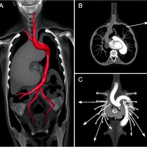
In a striking reminder of how complex the human body’s plumbing can be when development goes subtly awry, surgeons and imaging specialists at Qinzhou First People’s Hospital in Guangxi, China, have reported the successful repair of an extraordinarily rare combination of congenital cardiopulmonary anomalies in a 15-year-old girl: a type 2A bridging bronchus, a pulmonary artery sling, and an atrial septal defect. The case, published in BMC Pediatrics, highlights not only the anatomical rarity of this triad but also the decisive role that multimodality imaging played in tailoring an operation that avoided an otherwise routine-but-risky additional procedure.
The patient arrived with a history that had puzzled clinicians for years. She suffered from refractory respiratory symptoms and, most alarmingly, exertional syncope—fainting during physical activity, a symptom that in an adolescent demands serious cardiovascular investigation. When the medical team led by Jianliang Liu and corresponding author Chengchang Liu of the hospital’s Department of Cardiothoracic Surgery put her through a comprehensive preoperative workup, the results revealed a constellation of defects so unusual that the combination has rarely, if ever, been documented in the medical literature.
Each of the three anomalies on its own is uncommon; together, they represent a formidable diagnostic and surgical challenge. A bridging bronchus is a rare airway malformation in which the right upper lobe bronchus originates anomalously—typically from the trachea or, in the type 2A variant relevant here, from an aberrant position relative to the bronchial tree’s usual branching pattern. In a normal lung, the right bronchus divides into three lobar branches serving the upper, middle, and lower lobes. In a bridging bronchus, the bronchus to the right lung’s middle and lower lobes arises from the left main bronchus and then crosses the mediastinum—bridging over from one side to the other—to supply the right lung. The result is an airway architecture that can mimic other malformations and is easily missed on conventional imaging.
The second anomaly, a pulmonary artery sling, is a vascular defect with direct mechanical consequences for the airway. In this condition, the left pulmonary artery does not arise normally from the main pulmonary artery trunk. Instead, it branches off the right pulmonary artery and then courses backward between the trachea and the esophagus to reach the left lung. This aberrant route means the vessel literally slings around the trachea as it travels, and in many patients it compresses the airway, producing chronic wheezing, stridor, recurrent infections, and respiratory distress that often begins in infancy. Pulmonary artery sling is, in fact, the only vascular anomaly known to be associated with tracheobronchial malformations—which helps explain why it so frequently travels with a bridging bronchus.
The third element of the triad, an atrial septal defect, is a hole in the wall separating the heart’s two upper chambers. It is the most common congenital cardiac anomaly of its kind, but in combination with the two airway and vascular defects above, it added a hemodynamic dimension to the case: shunting of blood between the atria can, over years, overload the right side of the heart and the pulmonary circulation, contributing to symptoms such as exercise intolerance and fainting.
Diagnosis in such cases demands more than a single test, and the Chinese team deployed a trio of complementary imaging modalities, each of which contributed a distinct layer of anatomical information. Echocardiography, the workhorse of congenital heart evaluation, confirmed the atrial septal defect and characterized the intracardiac hemodynamics. Multi-detector computed tomography with three-dimensional reconstruction provided the pivotal contribution: a detailed, spatially accurate map of the aberrant airway and vascular anatomy that clarified exactly how the left pulmonary artery originated from the right pulmonary artery and how the bridging bronchus crossed the mediastinum. Bronchoscopy then allowed direct visual inspection of the tracheobronchial tree from within, corroborating the CT findings and, critically, assessing the caliber of the airway lumen itself.
That last point proved to be the linchpin of the entire surgical strategy. Pulmonary artery sling is famously associated with complete tracheal rings and significant tracheal stenosis—narrowing of the windpipe—which, when present, typically forces surgeons to perform a tracheoplasty in addition to correcting the vascular anomaly. Tracheoplasty substantially extends operative time, adds complexity, and carries its own risks of airway complications during recovery. Here, however, the imaging told a different and more favorable story: despite the anomalous bronchial architecture, the girl’s trachea showed no significant stenosis. The airway, though abnormally built, was not dangerously narrowed. That single imaging finding obviated the need for tracheoplasty entirely, allowing the team to plan a more streamlined operation addressing only the vascular and cardiac defects.
Armed with this anatomical roadmap, the surgical team proceeded with a repair conducted through a median sternotomy—the vertical incision through the breastbone that provides full access to the heart and great vessels—performed while the patient was supported by cardiopulmonary bypass, the heart-lung machine that temporarily takes over circulation and oxygenation. The operation had two principal components. First, the left pulmonary artery was detached from its anomalous origin off the right pulmonary artery and reimplanted in its correct position arising from the main pulmonary artery, eliminating the sling around the trachea and restoring normal vascular geometry. Second, the atrial septal defect was closed with a patch, sealing the abnormal communication between the atria and abolishing the pathological left-to-right shunt.
The outcome validated the preoperative planning. Postoperative multi-detector CT confirmed that the reconstructed left pulmonary artery was patent—open and carrying blood normally to the left lung—and the patient’s symptoms resolved. The fainting episodes that had brought her to medical attention disappeared, and the refractory respiratory complaints that had marked her earlier years were relieved once the compressive vascular sling was untangled and the cardiac shunt was closed.
The case carries broader lessons for pediatric cardiology, radiology, and congenital surgery. First, it underscores that bridging bronchus and pulmonary artery sling should be considered together whenever either is found, since their embryological origins intertwine during airway and vascular development. Patients presenting with unexplained, persistent respiratory symptoms—even in adolescence, long past the infancy window in which these anomalies usually declare themselves—may harbor anatomy that only advanced imaging can reveal. Second, the case demonstrates why three-dimensional CT reconstruction has become indispensable in this field: two-dimensional slices can suggest anomalies, but only volumetric rendering makes the spatial relationships of vessels and airways unambiguous enough to plan a definitive repair. Third, and perhaps most importantly, the findings show that imaging must directly interrogate the question that shapes the operation: whether tracheal stenosis exists. In this patient, the answer determined that a major additional procedure could be safely skipped, reducing surgical risk without compromising the outcome.
The report also illustrates the unusual natural history such anomalies can follow. Many patients with pulmonary artery sling present in infancy with severe respiratory compromise, but this girl reached her mid-teens before exertional syncope forced definitive evaluation. That delayed presentation likely reflects the absence of severe tracheal compression—consistent with the finding of no significant stenosis—allowing her to compensate for years despite the anatomical burden. It is a caution against assuming that congenital vascular rings and slings always announce themselves in the first months of life.
The team reports that the study was conducted in accordance with the Declaration of Helsinki, with the ethics requirement waived by Qinzhou First People’s Hospital for this retrospective case report, and written informed consent obtained from the patient’s legal guardians for both surgery and publication. The work received no specific external funding, and the authors declare no competing interests. The article was published as an open-access case report in BMC Pediatrics, received by the journal in October 2025 and accepted in August 2026.
For clinicians, the message distilled from this single teenager’s case is clear: when the airway and the pulmonary vasculature appear anomalous in combination, no single imaging study suffices, and no surgical plan should be finalized until the trachea’s integrity has been definitively assessed. Echocardiography, multi-detector CT with three-dimensional reconstruction, and bronchoscopy together formed a diagnostic chain in which each link informed the next, culminating in an operation that was neither more nor less extensive than the anatomy required. In the realm of rare congenital malformations, that precision—the ability to reimplant a pulmonary artery and patch an atrial septum while deliberately leaving the airway untouched—is what modern multimodality imaging makes possible.
Subject of Research: Surgical management, guided by multimodality imaging, of a rare combination of type 2A bridging bronchus, pulmonary artery sling, and atrial septal defect in a 15-year-old patient
Subject of Research: Medicine
Article Title: Multimodality imaging and surgical management of a rare combination: type 2A bridging bronchus, pulmonary artery sling, and atrial septal defect, a case report
Article References: Liu, J., Song, J., Pei, X., Li, C., Li, D., Lu, Y., Chen, J., & Liu, C. (2026). Multimodality imaging and surgical management of a rare combination: type 2A bridging bronchus, pulmonary artery sling, and atrial septal defect, a case report. BMC Pediatrics. https://doi.org/10.1186/s12887-026-07605-9
Image Credits: AI Generated
DOI: 10.1186/s12887-026-07605-9
Keywords: bridging bronchus, pulmonary artery sling, atrial septal defect, multi-detector computed tomography, congenital heart surgery, tracheal stenosis, case report
Cite Scienmag News
APA MLA Chicago
Ophelia Keating. (September 4, 2026). Imaging and surgery for rare bridging bronchus, artery sling, and heart defect. Scienmag. https://scienmag.com/imaging-and-surgery-for-rare-bridging-bronchus-artery-sling-and-heart-defect/
Ophelia Keating. “Imaging and surgery for rare bridging bronchus, artery sling, and heart defect.” Scienmag, 4 September 2026, https://scienmag.com/imaging-and-surgery-for-rare-bridging-bronchus-artery-sling-and-heart-defect/. Accessed 4 September 2026.
Ophelia Keating. “Imaging and surgery for rare bridging bronchus, artery sling, and heart defect.” Scienmag. September 4, 2026. https://scienmag.com/imaging-and-surgery-for-rare-bridging-bronchus-artery-sling-and-heart-defect/
Copy citation Download RIS
Tags: advanced imaging techniques in congenital anomaly assessmentadvances in imaging-guided congenital anomaly repairanatomical variations in respiratory and cardiovascular systematrial septal defect surgerybridging bronchus in childrencardiopulmonary anomalies diagnosiscase report of rare triad congenital anomaliescase report on congenital lung and heart defectschallenges in diagnosing and treating complex congenital heart defectscomplex airway and vascular malformationscongenital atrial septal defectCongenital cardiopulmonary anomaliesdiagnostic imaging for congenital anomaliesmultimodality imaging in congenital heart defectsmultimodality imaging in congenital heart diseasepediatric cardiothoracic surgerypediatric cardiothoracic surgery case studypulmonary artery slingpulmonary artery sling repairrare bridging bronchusrare thoracic vascular anomaliessurgical management of bridging bronchus and artery slingsurgical management of rare bronchial and arterial anomaliessurgical repair of complex congenital defects